Figure 8. Subcellular localization of p27 in the RPE. A: Immunofluorescence for p27 in the wild-type (WT) and p27 knockout (KO) retinas. Note p27 labeling in the outer nuclear layer
(ONL), inner nuclear layer (INL), and ganglion cell layer (GCL) in the WT retina and the absence of staining in the knockout
(KO) retina. B: Double immunofluorescence for p27 and Sox9 in the RPE of the WT and p27 KO retinas. Arrows indicate the Sox9-positive RPE
nuclei. Note the nuclear expression of p27 in the WT RPE and the absence of p27 in the mutant. Arrowheads denote Bruch’s membrane
labeled non-specifically. C: Double immunofluorescence for p27 and Sox9 in the WT RPE at day 5 after N-methyl-N-nitrosourea (MNU) treatment. p27 is localized
not only in the nuclei (arrows) but also in the cytoplasm (arrowheads). D: Double immunofluorescence for p27 and myosin VIIa (MyoVIIa) in the WT RPE at day 5 after MNU treatment. Note nuclear (arrows)
and cytoplasmic (arrowheads) staining for p27. DIC, differential interference contrast. Scale bars = 20 μm.
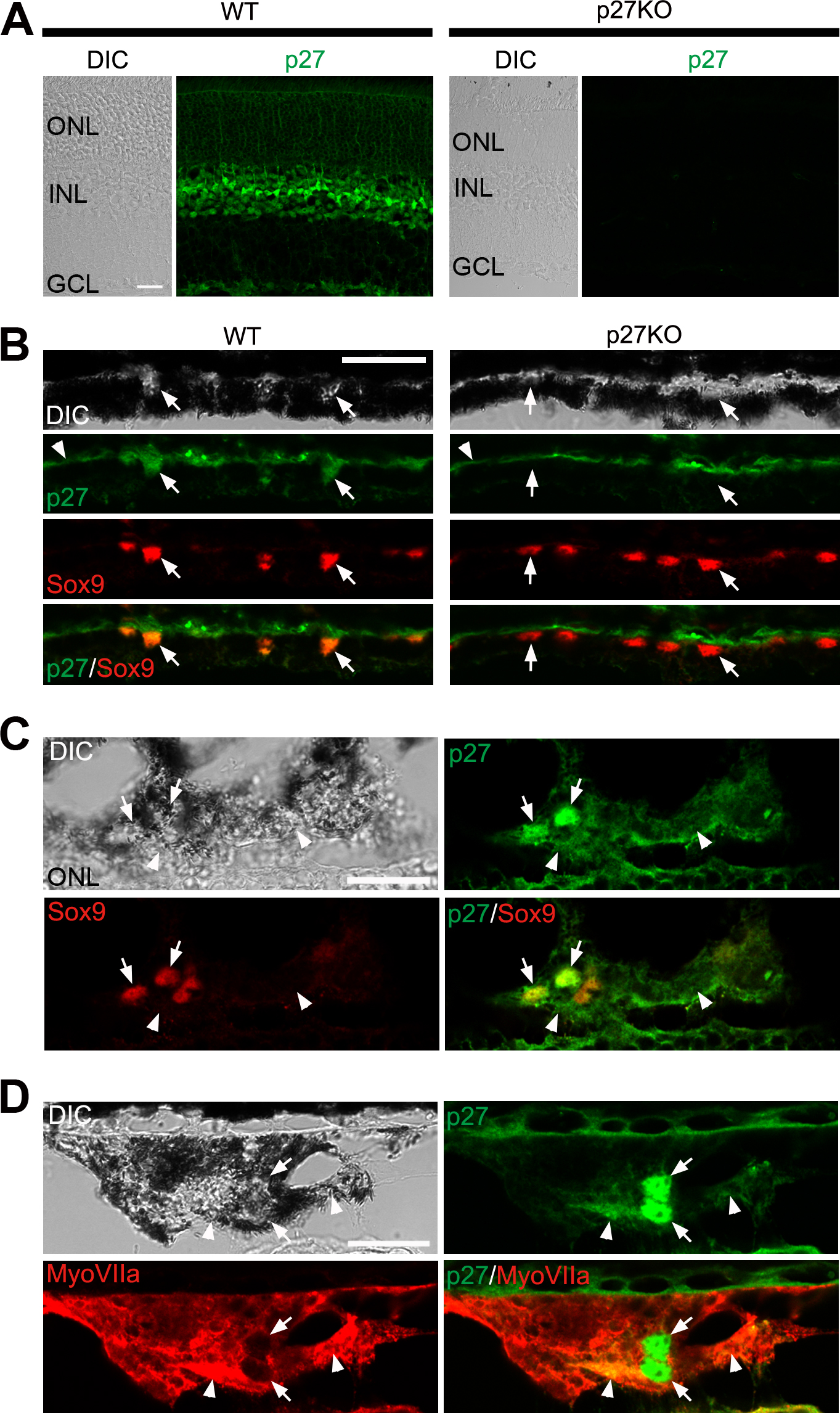
 Figure 8 of
ul Quraish, Mol Vis 2016; 22:1103-1121.
Figure 8 of
ul Quraish, Mol Vis 2016; 22:1103-1121.