Figure 5. Enhanced phosphorylation of myosin light chains in the RPE of the p27 KO retinas after photoreceptor damage. A: Immunofluorescence for phosphorylated myosin light chains (pMLC) combined with phalloidin labeling. Arrows indicate colocalization
of phalloidin-labeled F-actin and pMLC in the apical junctions of the RPE. The outer limiting membrane is also labeled for
F-actin (arrowheads). Note enhanced labeling for F-actin and pMLC in the mutant RPE at day 3. B: Triple labeling for F-actin, pMLC, and rhodopsin (Rho) in the mutant RPE at day 3. Note colocalization of F-actin and pMLC
with Rho-positive phagosomes (arrows). DIC, differential interference contrast; Cont, control; OS, outer segments; ONL, outer
nuclear layer; INL, inner nuclear layer. Scale bars = 20 μm.
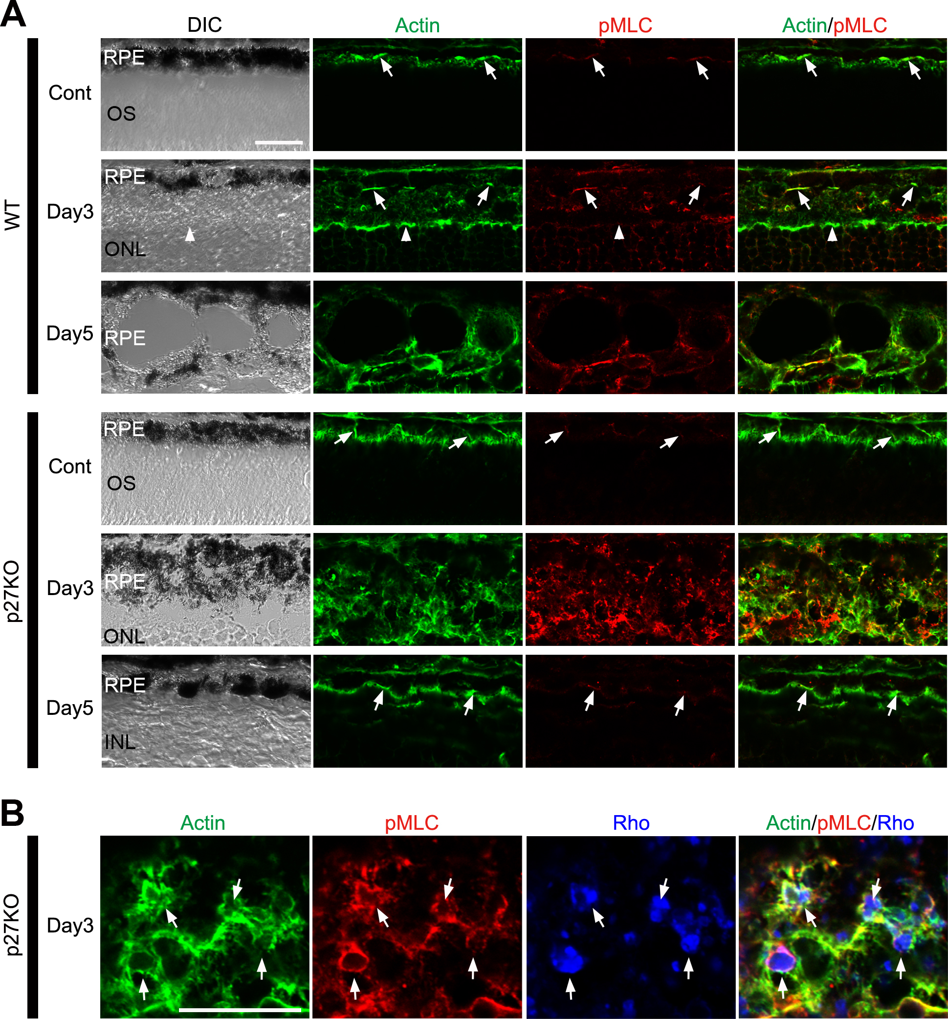
 Figure 5 of
ul Quraish, Mol Vis 2016; 22:1103-1121.
Figure 5 of
ul Quraish, Mol Vis 2016; 22:1103-1121.