Figure 3. Enhanced phagocytosis of photoreceptor debris by the RPE in the p27 KO retinas. A: Rhodopsin immunofluorescence displaying the outer nuclear layer (ONL) and photoreceptor outer segments. Dotted lines indicate
the thickness of the ONL. Arrows denote intensely labeled outer segment debris in the wild-type (WT) retina. Cont, control.
Scale bar = 20 μm. B: Quantitative analysis of the areas of the ONL expressed as percentages relative to the value in the intact WT retina. Each
bar represents the mean ± standard error of the mean (SEM; n = 3). Asterisks denotes statistically significant difference
(p<0.01). C: Double immunofluorescence for myosin VIIa (MyoVIIa) and rhodopsin (Rho) showing an accumulation of Rho-positive phagosomes
(arrowheads) in the RPE after photoreceptor damage. Engulfment of Rho-positive photoreceptor cell bodies (arrows) by the RPE
is found only in the knockout (KO) retinas. Terminal deoxynucleotidyl transferase dUTP nick end labeling (TUNEL) assays combined
with MyoVIIa labeling reveal dead photoreceptor nuclei (arrows) engulfed by the RPE in the KO retinas. OS, outer segments;
INL, inner nuclear layer; ONL, outer nuclear layer. Scale bars = 20 μm. D: Nuclear staining of the whole mount RPE with Hoechst 33258 showing photoreceptor nuclei (arrows) engulfed by the mutant
RPE at day 3 after N-methyl-N-nitrosourea (MNU) treatment. Arrowheads indicate the RPE nuclei.
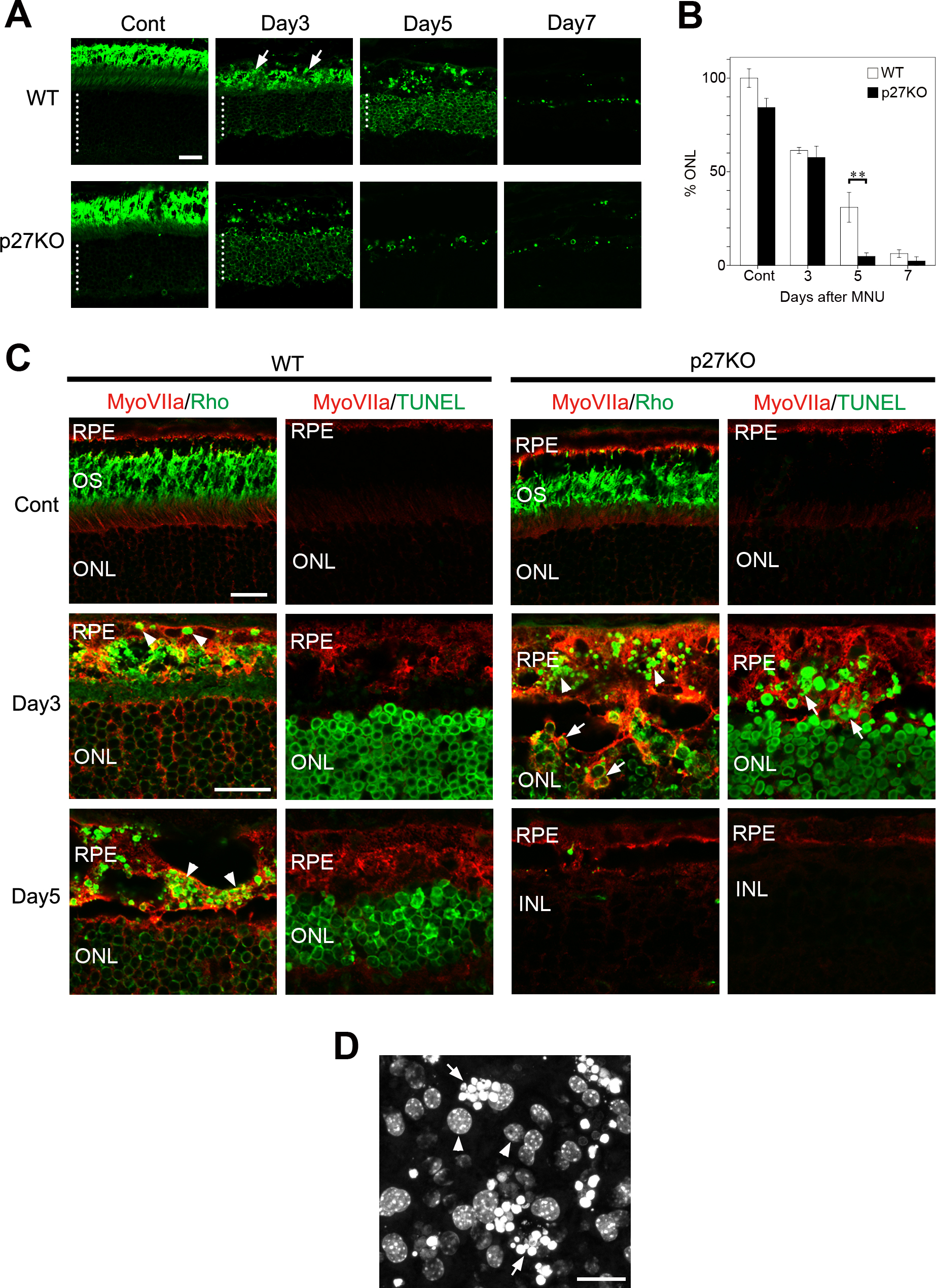
 Figure 3 of
ul Quraish, Mol Vis 2016; 22:1103-1121.
Figure 3 of
ul Quraish, Mol Vis 2016; 22:1103-1121.