Figure 2. Myosin VIIa immunofluorescence in the RPE of the WT and p27 KO retinas after MNU treatment. The wild-type (WT) RPE becomes
thicker and vacuolated (asterisks) by day 5 after MNU treatment. The RPE in the knockout (KO) retinas develops many apical
protrusions as early as day 1. Arrows indicate RPE protrusions in the mutant retina invading into the outer nuclear layer
(ONL) at day 3. DIC, differential interference contrast; MyoVIIa, myosin VIIa; Cont, control; INL, inner nuclear layer. Scale
bar = 20 μm.
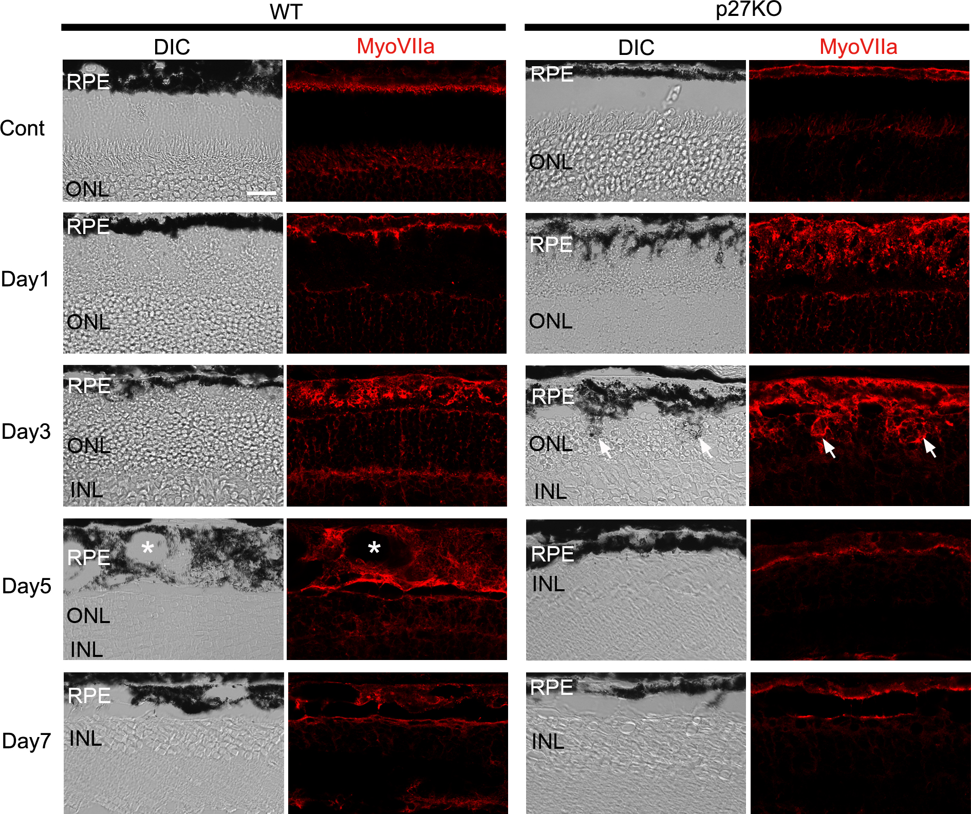
 Figure 2 of
ul Quraish, Mol Vis 2016; 22:1103-1121.
Figure 2 of
ul Quraish, Mol Vis 2016; 22:1103-1121.