Figure 1. Proliferation of Müller glia and RPE cells in the WT and p27 KO retinas after MNU-induced photoreceptor damage. A: Terminal deoxynucleotidyl transferase dUTP nick end labeling (TUNEL) assays. In the wild-type (WT) and knockout (KO) retinas,
most photoreceptor cells in the outer nuclear layer (ONL) are labeled at day 2 after MNU treatment while no labeling is observed
in the non-treated control retinas (Cont). B: Double immunofluorescence for Sox9 and bromodeoxyuridine (BrdU). Note BrdU-/Sox9-positive Müller glia (arrows) and RPE cells
(arrowheads) in the KO retinas at day 2. Virtually no BrdU-positive Müller glia or RPE cells are observed in the WT retinas.
C: The proportion of BrdU-positive Müller glia and RPE cells in the central retina. Each bar represents the mean ± standard
error of the mean (SEM; n = 3). Asterisks denote statistically significant differences between the WT and KO retinas (*p<0.05;
**p<0.01). DIC, differential interference contrast; INL, inner nuclear layer; GCL, ganglion cell layer. Scale bars = 20 μm.
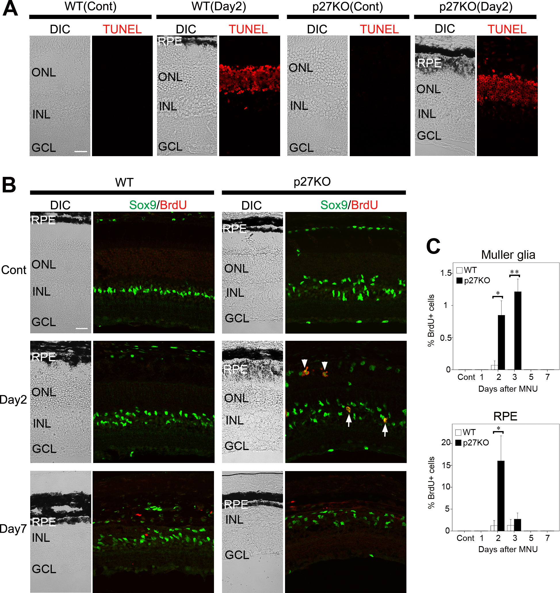
 Figure 1 of
ul Quraish, Mol Vis 2016; 22:1103-1121.
Figure 1 of
ul Quraish, Mol Vis 2016; 22:1103-1121.