Figure 3. Corneal thickness after therapy. Whole eye tissue sections were stained with hematoxylin and eosin (H&E) to measure the corneal
epithelial thickness. The epithelial layer is shown in white brackets with a representative thickness measurement. A: Normal eye. B: Cyclosporine. C: Dry eye, no therapy. D: Dry eye treated with whole milk, 10X magnification, red bar = 100 μm.
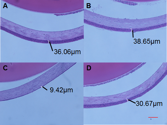
 Figure 3 of
Diego, Mol Vis 2016; 22:1095-1102.
Figure 3 of
Diego, Mol Vis 2016; 22:1095-1102.