Figure 8. Maturation of photoreceptors and synaptic layers in the retinal organoids (day (D)25–32). No significant morphological differences
were observed among the neural retinas differentiated from embryonic pluripotent stem cells (ESCs) or induced pluripotent
stem cells (iPSCs); representative figures of the neural retinas are shown. Nuclei were stained with 4',6-diamidino-2-phenylindole
(DAPI; blue). A: Appearance of photoreceptor cilia. Gamma-tubulin (γ-tubulin, red) is a marker of basal bodies, whereas ADP-ribosylation
factor-like protein 13B (Arl13b, green) stains the ciliary axoneme of photoreceptors. B: Labeling of rhodopsin (Rho, green) beyond Arl13b (red) staining. C: Synaptophysin (green) labeling indicates the synaptic layers. The white rectangles show higher magnification images of the
selected region. AP and BA show the apical and basal sides of the organoid, respectively. NBL, neuroblastic layer; ONL, outer
nuclear layer; INL, inner nuclear layer; GCL, ganglion cell layer. Scale bar = 10 μm.
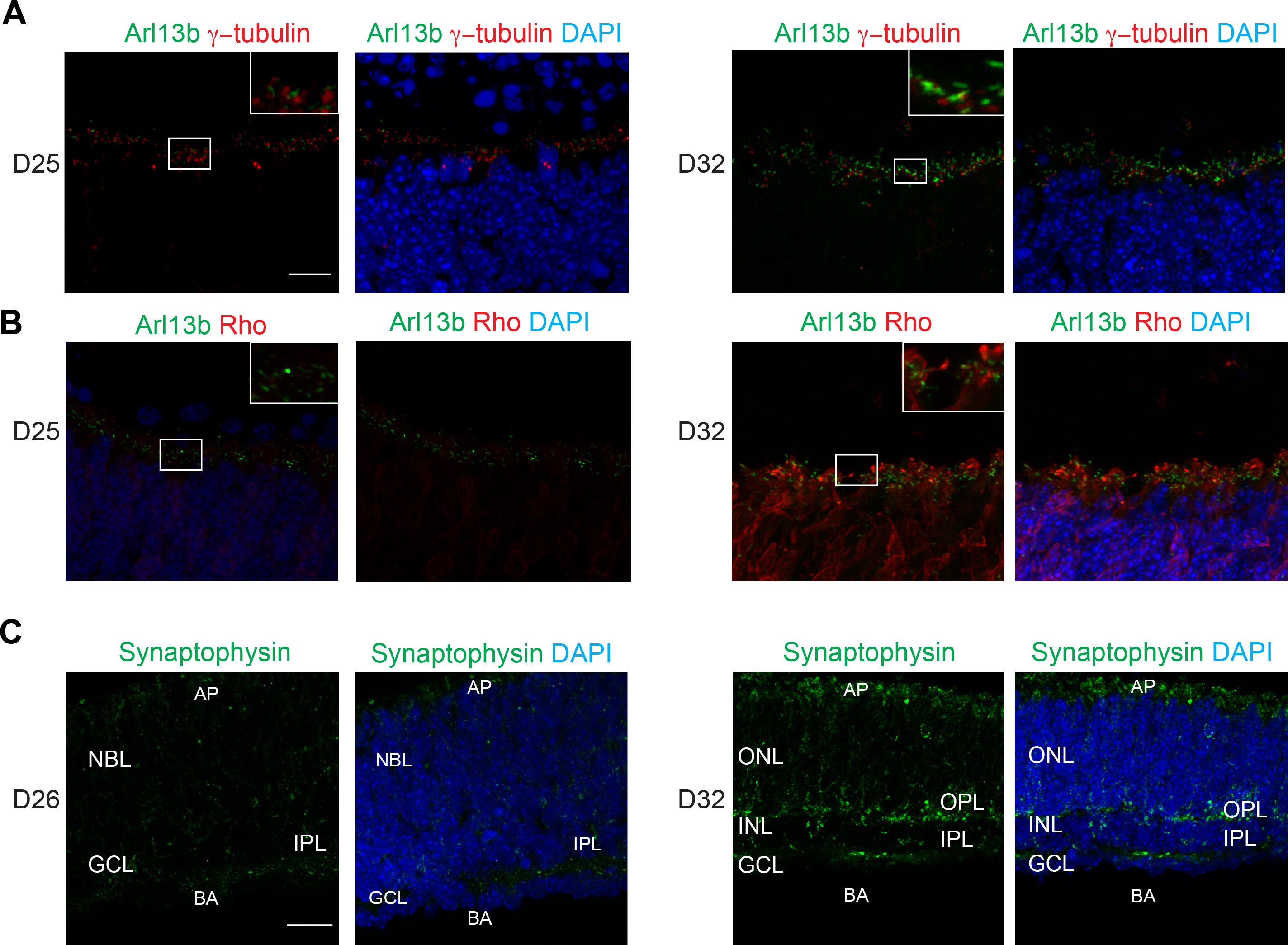
 Figure 8 of
Chen, Mol Vis 2016; 22:1077-1094.
Figure 8 of
Chen, Mol Vis 2016; 22:1077-1094.