Figure 7. Differentiation of rod photoreceptors and Müller glia in developing retinal organoids (day (D)18–35). Reep6 (green) and glutamine
synthetase (GS, red) are markers of rod photoreceptors and Müller glia, respectively. No significant morphological differences
were observed among the neural retinas differentiated from embryonic pluripotent stem cells (ESCs) or induced pluripotent
stem cells (iPSCs); representative figures of the neural retinas are shown. Nuclei were stained with 4',6-diamidino-2-phenylindole
(DAPI; blue). Arrowheads indicate the relevant immunostaining with Reep6 and GS. AP and BA show the apical and basal sides
of the organoid, respectively. NBL, neuroblastic layer; ONL, outer nuclear layer; INL, inner nuclear layer; GCL, ganglion
cell layer. Scale bar = 50 μm.
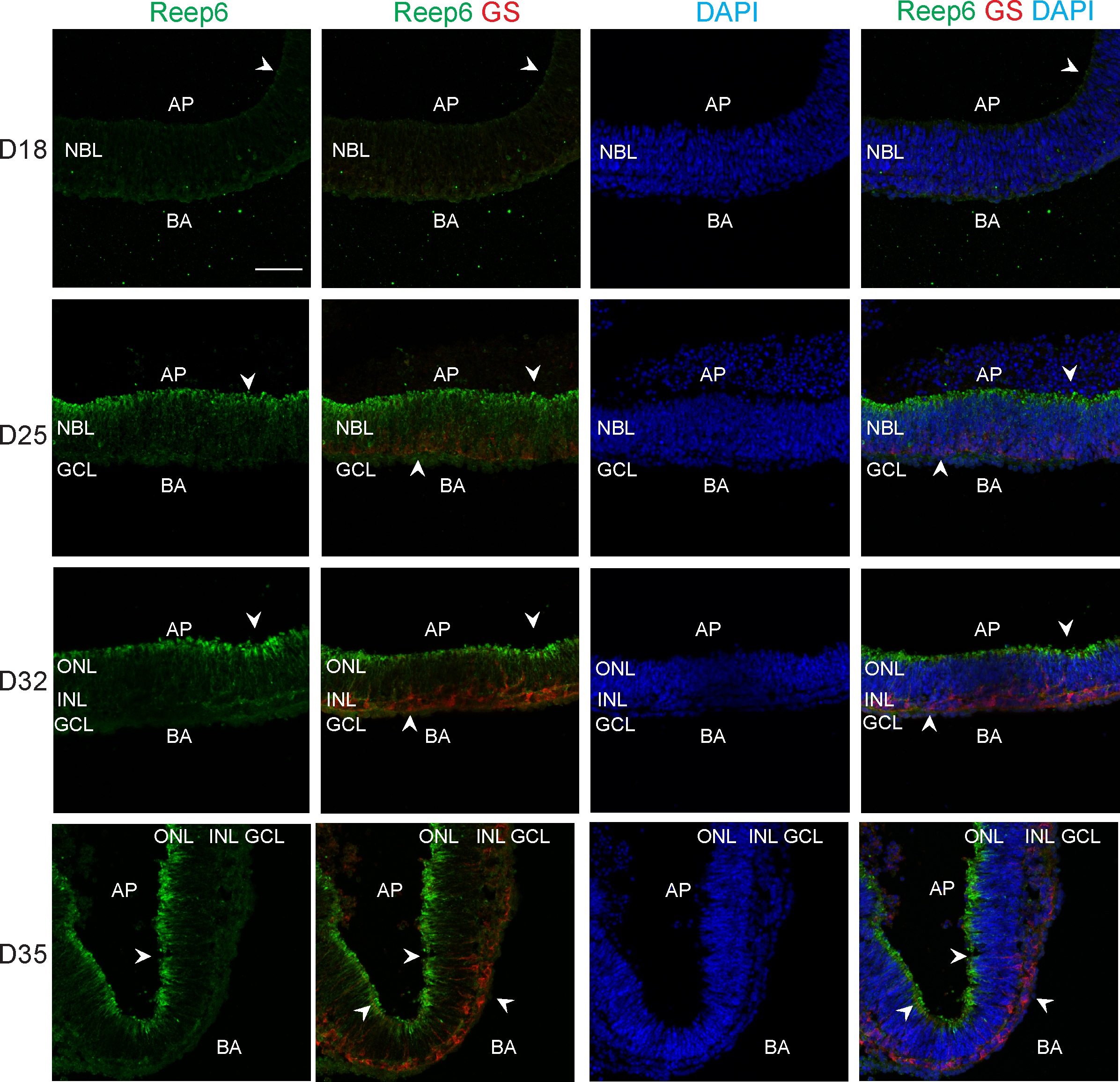
 Figure 7 of
Chen, Mol Vis 2016; 22:1077-1094.
Figure 7 of
Chen, Mol Vis 2016; 22:1077-1094.