Figure 5. Genesis of bipolar cells in retinal organoids at different stages of differentiation (day (D)18 to D32). Ceh-10 homeodomain-containing
homolog (Chx10; red) is a marker of proliferating cells as well as differentiated bipolar cells. Protein kinase C alpha (PKCα;
green) is a marker of ON-bipolar cells. No significant morphological differences were observed among the neural retinas differentiated
from embryonic pluripotent stem cells (ESCs) or induced pluripotent stem cells (iPSCs); representative figures are shown.
Nuclei were stained with 4',6-diamidino-2-phenylindole (DAPI; blue). Arrowheads indicate the relevant immunostaining in the
inner retina. At D18 and D26, the Chx10 labeling shows mostly proliferating cells. AP and BA show the apical and basal sides
of the organoid, respectively. NBL, neuroblastic layer; ONL, outer nuclear layer; INL, inner nuclear layer; GCL, ganglion
cell layer. Scale bar = 50 μm.
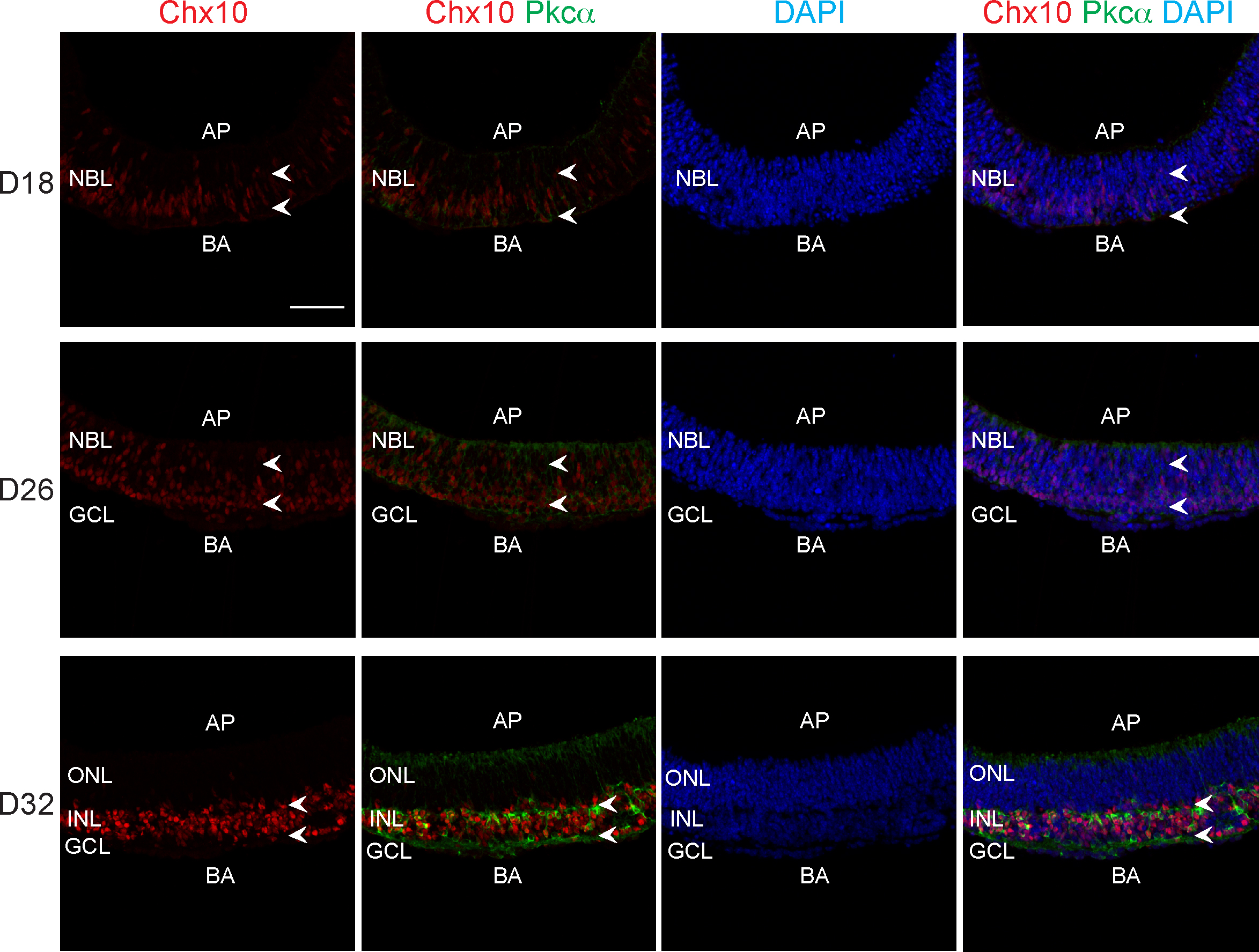
 Figure 5 of
Chen, Mol Vis 2016; 22:1077-1094.
Figure 5 of
Chen, Mol Vis 2016; 22:1077-1094.