Figure 4. Development of cone photoreceptors and amacrine cells, followed by the formation of plexiform layers. Immunostaining with
S-opsin (Opnsw, red) and Prox1 (green) antibodies shows S-cones and amacrine cells, respectively. No significant morphological
differences were observed among the neural retinas differentiated from embryonic pluripotent stem cells (ESCs) or induced
pluripotent stem cells (iPSCs); representative figures are shown. Arrowheads indicate the relevant immunostaining with Opnsw
and Prox1. AP and BA show the apical and basal sides of the organoid, respectively. NBL, neuroblastic layer; ONL, outer nuclear
layer; INL, inner nuclear layer; GCL, ganglion cell layer. Nuclei were stained with 4',6-diamidino-2-phenylindole (DAPI; blue).
Scale bar = 50 μm.
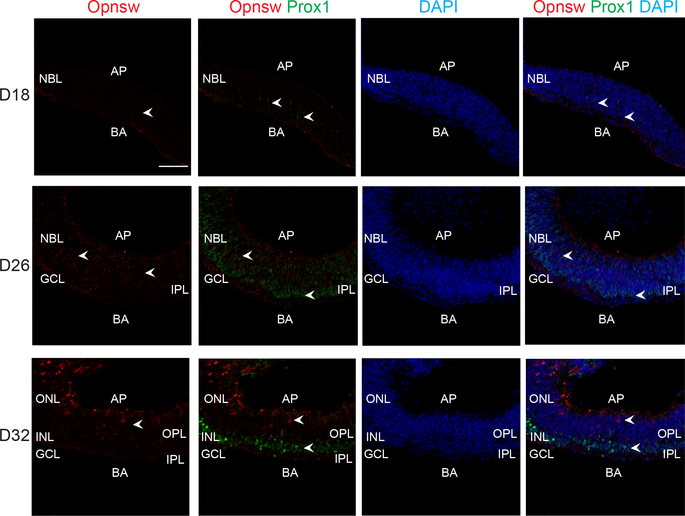
 Figure 4 of
Chen, Mol Vis 2016; 22:1077-1094.
Figure 4 of
Chen, Mol Vis 2016; 22:1077-1094.