Figure 3. Differentiation of early-born cell types in the polarized neural retina. AP and BA show the apical and basal sides of the
organoid, respectively. Nuclei were stained with 4',6-diamidino-2-phenylindole (DAPI; blue). No significant morphological
differences were observed among the neural retinas differentiated from the embryonic pluripotent stem cells (ESCs) or induced
pluripotent stem cells (iPSCs); representative figures are shown. Arrowheads indicate relevant immunostaining with organoid
polarity in (A), and Brn3a and Calb in (B). A: Polarity of the three-dimensional (3D) retina at day (D)14 and D25. Laminin (red) and ZO-1 (red) are basal and apical markers,
respectively. B: Development of ganglion cells (Brn3a, green) and horizontal cells (Calbindin, Calb, red) in the D14 to 21 retinal organoids.
NBL, neuroblastic layer; ONL, outer nuclear layer; INL, inner nuclear layer; GCL, ganglion cell layer. Scale bar = 50 μm.
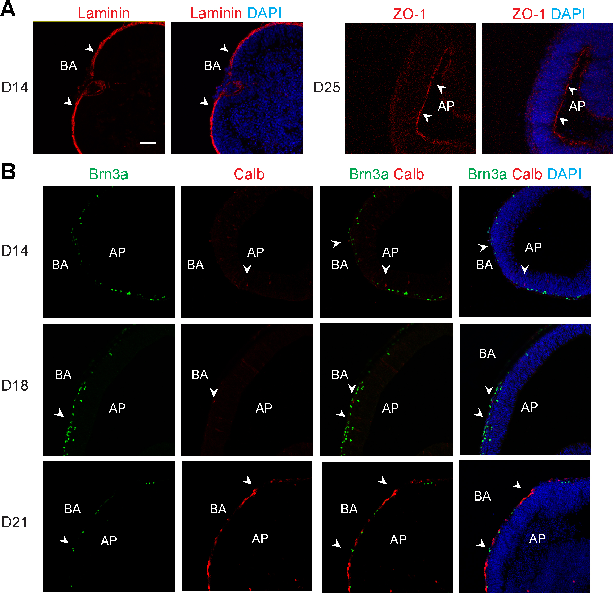
 Figure 3 of
Chen, Mol Vis 2016; 22:1077-1094.
Figure 3 of
Chen, Mol Vis 2016; 22:1077-1094.