Figure 2. Generation of retinal organoids from Nrl-GFP mouse ESCs and iPSCs. A: Schematic representation of the High Efficiency Hypoxia Induced Generation of Photoreceptors in Retinal Organoids (HIPRO)
protocol for differentiation of mouse stem cells into the three-dimensional (3D) neural retina. GMEM, Glasgow minimum essential
medium; KSR, knockout serum replacement; poly-HEMA, polyhydroxyethylmethacrylate; RMM, retinal maturation medium constituted
of DMEM/F12 with GlutaMAX, 1X penicillin-streptomycin, and 1X N2 supplement; RA, retinoic acid; FBS, fetal bovine serum; N2, N2 supplement; B27, B27 supplement. B: Morphology of organoids at different time points using normoxic and hypoxic conditions. C: Percentage of organoids that form optic vesicles at day (D)7 and optic cups at D10. Optic vesicles are defined as protrusions
of neuroepithelia from the organoids. Optic cups are characterized by a hinge region due to inward folding of the neuroepithelia
of the optic vesicles. The data were obtained from ten independent experiments (n = 10, each had a total of 144 organoids)
and represented as mean ± standard error of the mean (SEM); *p<0.05. D: Reverse transcription PCR (RT–PCR) analysis of early eye field transcription factors (Pax 6 and Rax) using total RNA from D10 organoids (60 organoids derived from embryonic pluripotent stem cells (ESCs) or 60 organoids from
induced pluripotent stem cells (iPSCs)). Gapdh was used as a control.
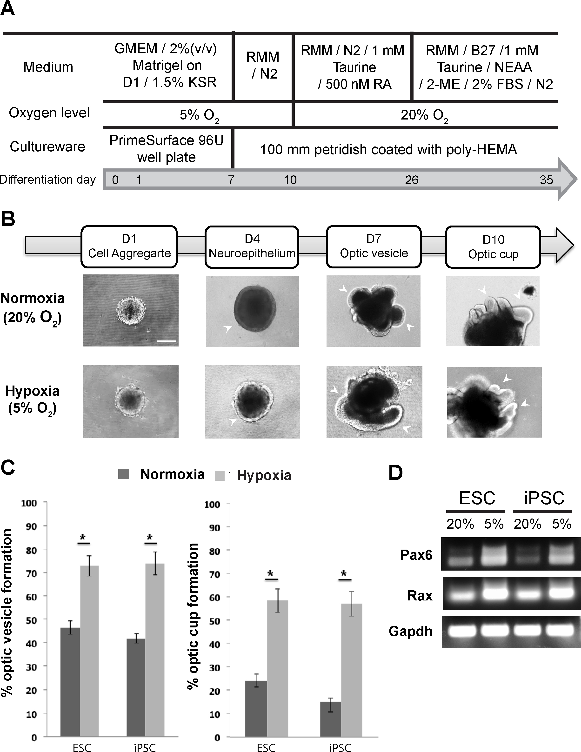
 Figure 2 of
Chen, Mol Vis 2016; 22:1077-1094.
Figure 2 of
Chen, Mol Vis 2016; 22:1077-1094.