Figure 1. Characterization of mouse ESCs and iPSCs. (A) morphology, (B) proliferation rate, (C) expression of pluripotency marker Nanog, and (D) percentage of Nanog+ cells. No significant morphological differences were observed between the embryonic pluripotent stem
cells (ESCs) and the induced pluripotent stem cells (iPSCs). Nanog staining is representative of both stem cell types. Nuclei
were stained with 4',6-diamidino-2-phenylindole (DAPI; blue). Scale bar = 100 μm.
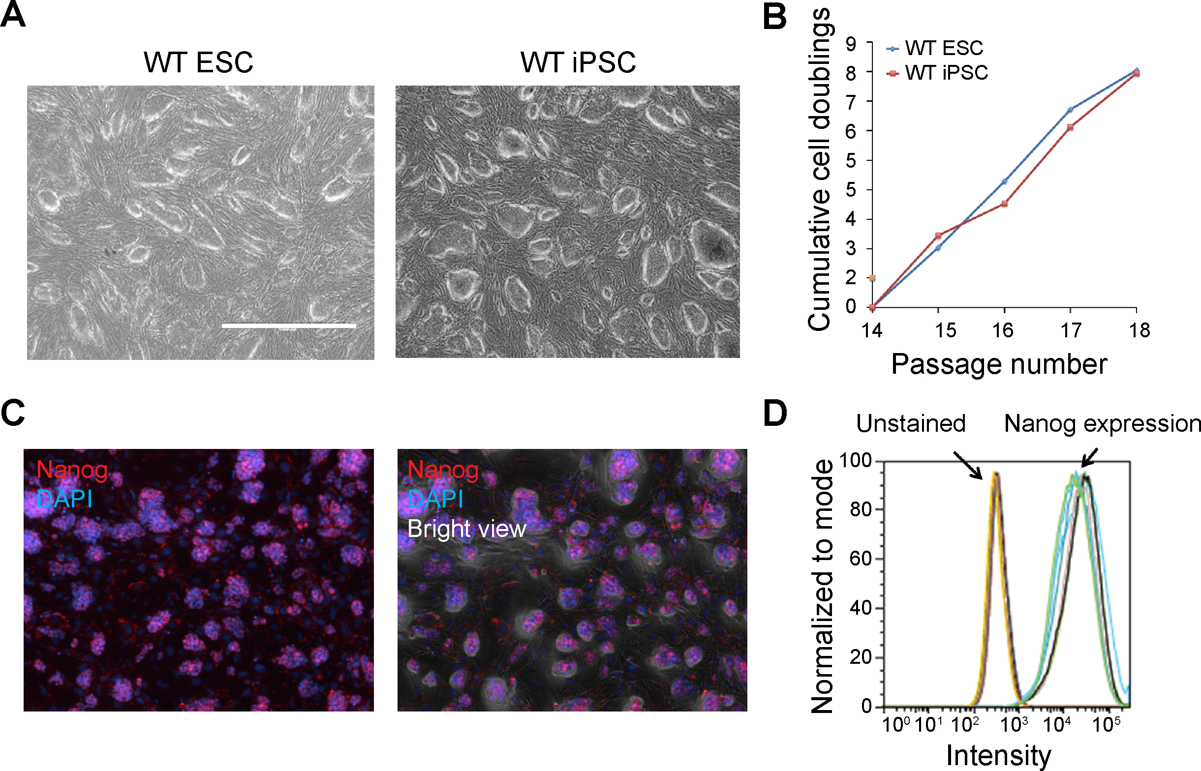
 Figure 1 of
Chen, Mol Vis 2016; 22:1077-1094.
Figure 1 of
Chen, Mol Vis 2016; 22:1077-1094.