Figure 6. Levels of p-AKT in the retinas of rats with elevated IOP overexpressing Brn3b. A: Immunostaining for p-AKT in retinal ganglion cells (RGCs) of Brown Norway rats intravitreally injected with either the recombinant
adenoassociated virus–cytomegalovirus–green fluorescent protein (rAAV-CMV-GFP) or rAAV-CMV-Brn3b following intraocular pressure
(IOP) elevation. Retinal sections obtained were immunostained for p-AKT (pseudogreen) and βIII-tubulin (pseudored). B: An increase in immunostaining (not statistically significant) for p-AKT was observed in RGCs overexpressing Brn3b (rAAV-CMV-Brn3b)
compared to RGCs overexpressing the control vector (rAAV-CMV-GFP; determined with the L/R ratios of fluorescence intensities
of p-AKT staining in RGCs). Values are represented as mean ± standard error of the mean (SEM), n = 3. Scale bar indicates
20 µm.
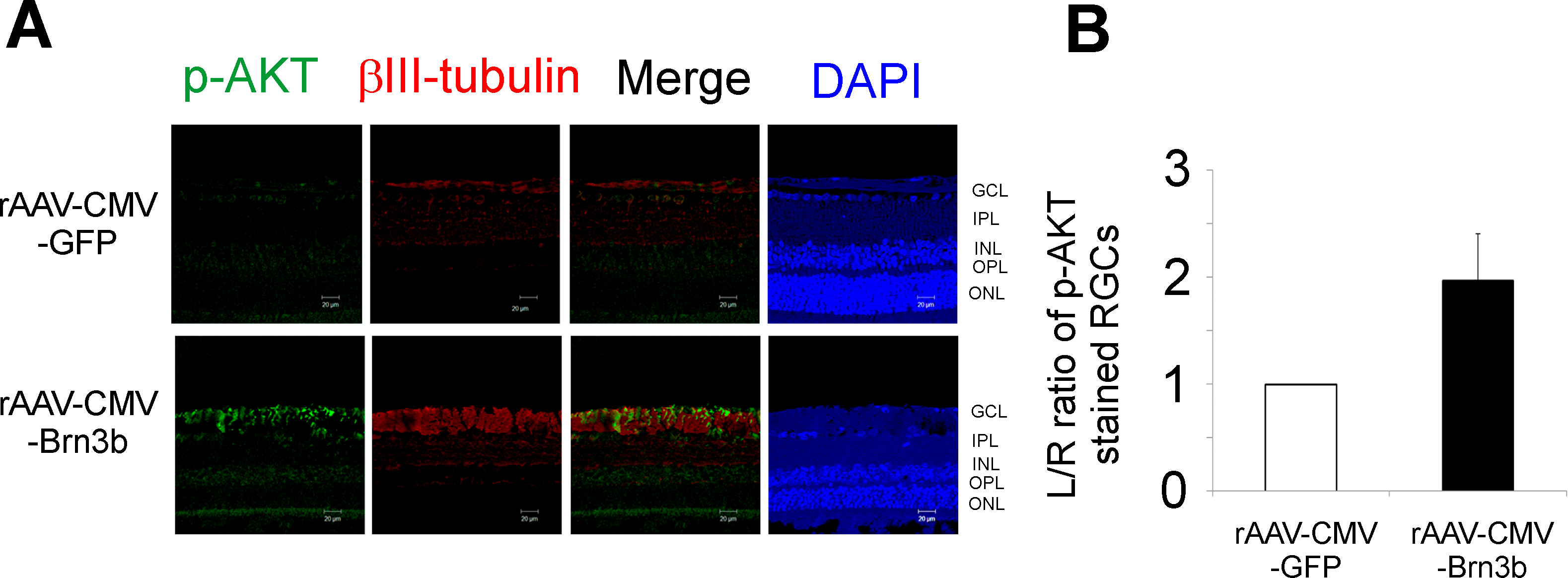
 Figure 6 of
Phatak, Mol Vis 2016; 22:1048-1061.
Figure 6 of
Phatak, Mol Vis 2016; 22:1048-1061.