Figure 4. AAV-mediated overexpression of Brn3b in RGCs of Brown Norway rats with elevated IOP following intravitreal injection of rAAV-CMV-Brn3b.
A: Representative images show Brn3b (pseudogreen) and βIII-tubulin (pseudored) immunostaining in retinas of rats intravitreally
injected with either recombinant adenoassociated virus–cytomegalovirus–green fluorescent protein (rAAV-CMV-GFP) or rAAV-CMV-Brn3b.
Brn3b staining was detected mainly in the GCL. B: Intraocular pressure (IOP) elevation profile in Brown Norway rats administered rAAV-CMV-GFP. C: IOP elevation profile in Brown Norway rats administered rAAV-CMV-Brn3b. IOP was elevated in one eye (closed circles), while
the other eye served as the contralateral control eye (open circles). IOP elevation was performed in three Brown Norway rats
followed by intravitreal injection with either rAAV-CMV-GFP or rAAV-CMV-Brn3b. IOP values were plotted as mean ± standard
error of the mean (SEM; solid line, IOP-elevated and virus-injected eye; dashed line, untreated contralateral eye). Asterisk
indicates p<0.001 statistically significant elevation of IOP in the IOP-elevated eye compared with contralateral eye using
the Student t test. Scale bar indicates 20 µm.
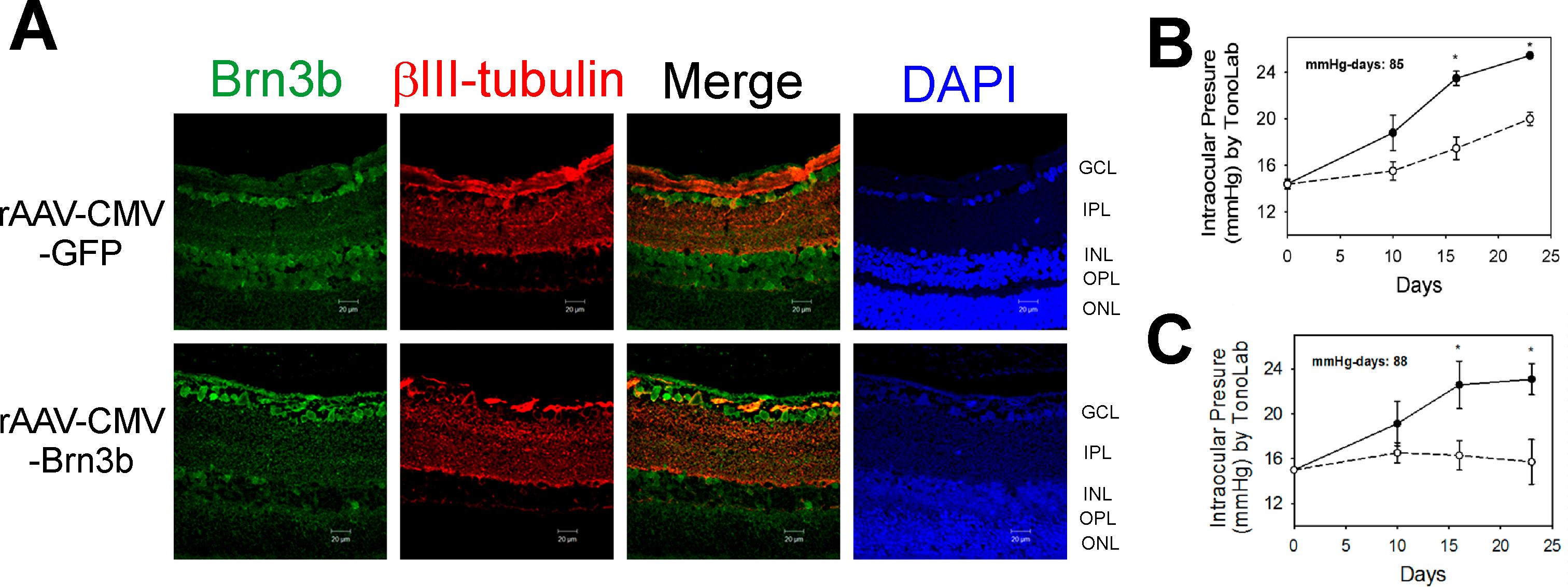
 Figure 4 of
Phatak, Mol Vis 2016; 22:1048-1061.
Figure 4 of
Phatak, Mol Vis 2016; 22:1048-1061.