Figure 2. Transcription factor Brn3b-mediated changes in Bcl-2 and Bcl-xL expression in RGCs of Brown Norway rats. A: Bcl-2 (pseudogreen), and βIII-tubulin (pseudored) expression in retinal sections from Brown Norway rat eyes injected with
either the recombinant adenoassociated virus–hSyn–green fluorescent protein (rAAV-hSyn-GFP) virus (control) or the rAAV-hSyn-Brn3b
virus, detected with secondary antibodies conjugated with Alexa 546 (pseudogreen) or Alexa 647 (pseudored) dye. Cells were
counterstained with 4',6-diamidino-2-phenylindole (DAPI; blue) to detect cell nuclei. B: Magnified image of the retinal ganglion cell (RGC) layers showing Bcl-2 immunostaining of RGCs of retinas transduced with
either rAAV-hSyn-GFP or rAAV-hSyn-Brn3b. Scale bar indicates 20 µm. C: Ratio of fluorescence intensity for Bcl-2 staining was measured at 24 different regions in the ganglion cell layers using
ImageJ. The fluorescence intensity ratios are shown as mean ± standard error of the mean (SEM), n = 6. A statistically significant
increase in the intensity of Bcl-2 staining was found in the RGCs of the rats injected with rAAV-hSyn-Brn3b, compared to those
injected with rAAV-hsyn-GFP. Statistical analysis was performed with the Mann–Whitney rank-sum test (*p<0.002). D: Bcl-xL (pseudogreen) and βIII-tubulin (pseudored) immunostaining in frozen retinal sections from rats intravitreally injected
with either rAAV-hSyn-GFP or rAAV-hSyn-Brn3b.
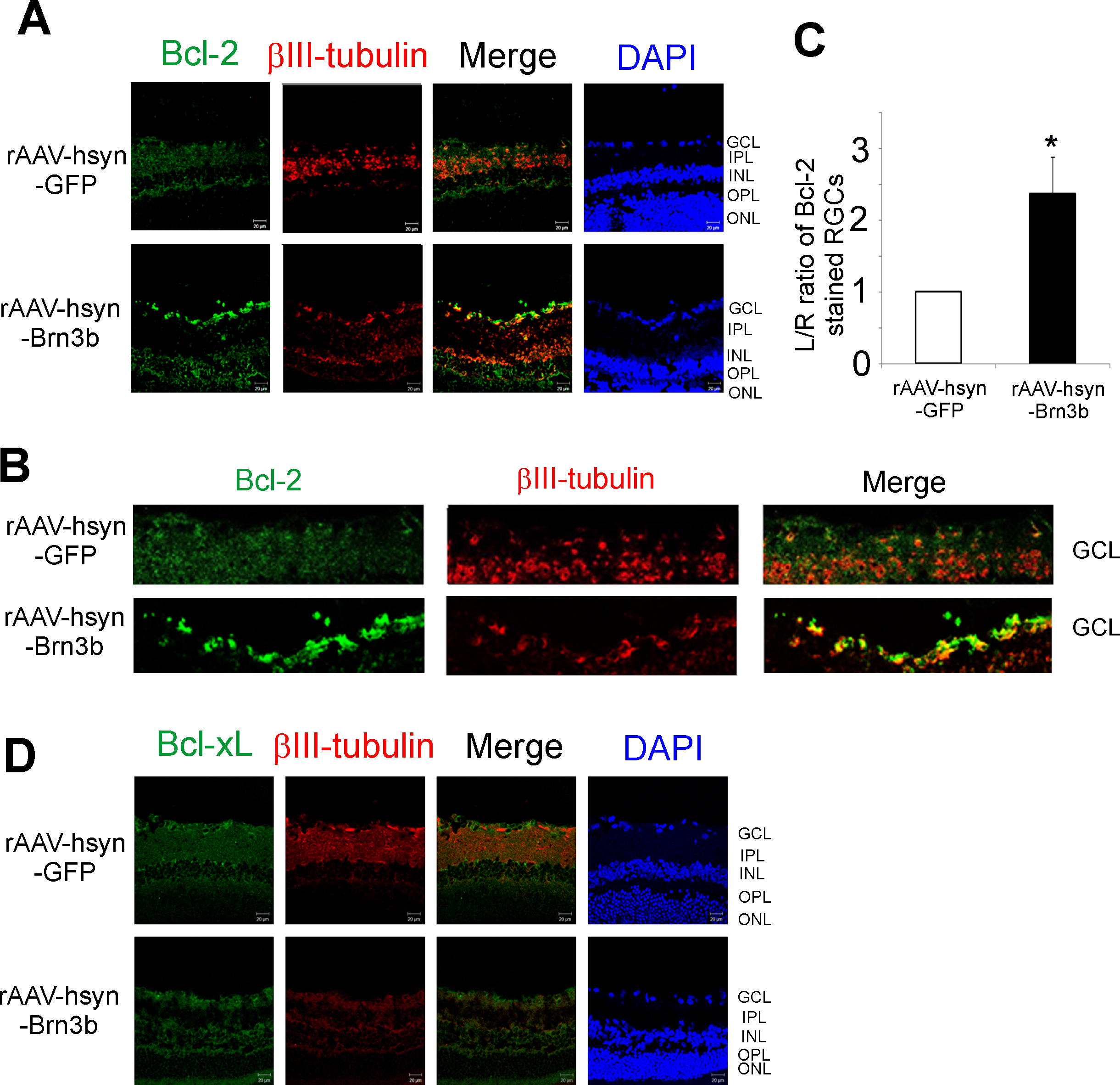
 Figure 2 of
Phatak, Mol Vis 2016; 22:1048-1061.
Figure 2 of
Phatak, Mol Vis 2016; 22:1048-1061.