Figure 4. SP activates the Akt/GSK-3β signaling in RPE cells under oxidative stress. RPE cells were treated with 200 μm H2O2 for 24 h, and substance P (SP) was added for 5, 10, and 15 min. A: The levels of phospho-Akt and phospho-glycogen synthase kinase (GSK)-3β were detected with western blotting. B, C: Phospho-Akt and phospho-GSK-3β protein expression levels, relative to the total Akt and GSK-3β, were quantified using the
Image J program. The expression level was represented relative to that of the untreated control. P values of less than 0.05
were considered statistically significant (*p<0.05, **p<0.01, ***p<0.001). The data are expressed as the mean ± standard deviation
(SD) of three independent experiments.
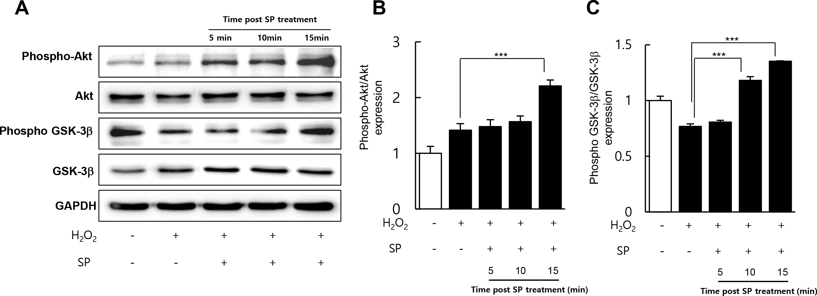
 Figure 4 of
Baek, Mol Vis 2016; 22:1015-1023.
Figure 4 of
Baek, Mol Vis 2016; 22:1015-1023.