Figure 3. SP increases cell viability by inhibiting apoptosis and promoting the proliferation of RPE cells damaged by oxidative stress.
RPE cells were damaged by H2O2, and then treated with substance P (SP) for 24 h. The effects of SP on cell apoptosis and proliferation were evaluated. A: The terminal deoxynucleotidyl transferase dUTP nick end labeling (TUNEL) assay was performed to quantify the number of apoptotic
RPE cells. B: The 5′-bromo-2′-deoxyuridine (BrdU) incorporation assay was performed to determine the proliferating cell pool. Representative
images for BrdU-incorporated RPE cells. Yellow arrow: BrdU (+) cells. C: BrdU (+) cells were quantified by counting BrdU (+) cells from the total PI (+) cells and expressing them as a percentage.
Scale bar: 100 μm. P values of less than 0.05 were considered statistically significant (*p<0.05, **p<0.01, ***p<0.001). The
data are expressed as the mean ± standard deviation (SD) of three independent experiments.
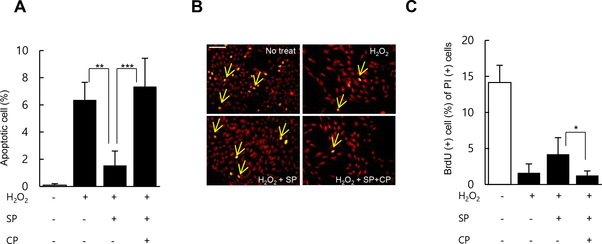
 Figure 3 of
Baek, Mol Vis 2016; 22:1015-1023.
Figure 3 of
Baek, Mol Vis 2016; 22:1015-1023.