Figure 3. Gamma-aminobutyric acidAα1 (GABAAα1) and GABAAρ1 protein expression in cultured human RPE cells detected by western blots (A) and immunofluorescence (B) (representative image;
n = 5). A: Specific bands presented at the approximate location of 51 kDa (GABAAα1), 48 kDa (GABAAρ1), and 43 kDa (β-Actin) in lysates of RPE and the retina (positive control, PC). B: Immunofluorescence staining of GABAAα1 and GABAAρ1 in cultured human RPE. Nuclei were stained by 4',6-diamidino-2-phenylindole (DAPI; blue; bar = 20 μm). NC, negative control.
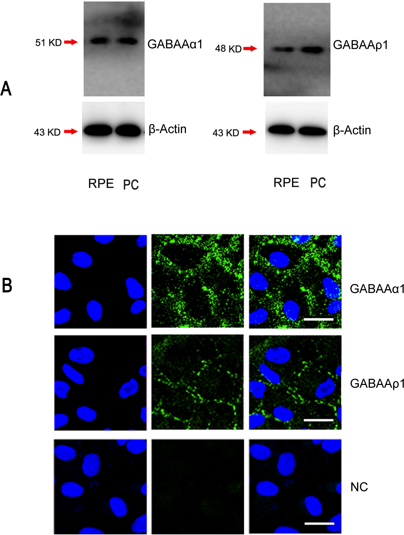
 Figure 3 of
Cheng, Mol Vis 2015; 21:939-947.
Figure 3 of
Cheng, Mol Vis 2015; 21:939-947.