Figure 1. Phenotype identification of the cultured human RPE cells using immunofluorescence (representative image; n = 5). A: All of the cultured cells were positively stained with the RPE65 antibody. B: All of the cultured cells were negatively stained with the S100 antibody. Nuclei were stained by 4',6-diamidino-2-phenylindole
(DAPI). Scale bar = 50 μm.
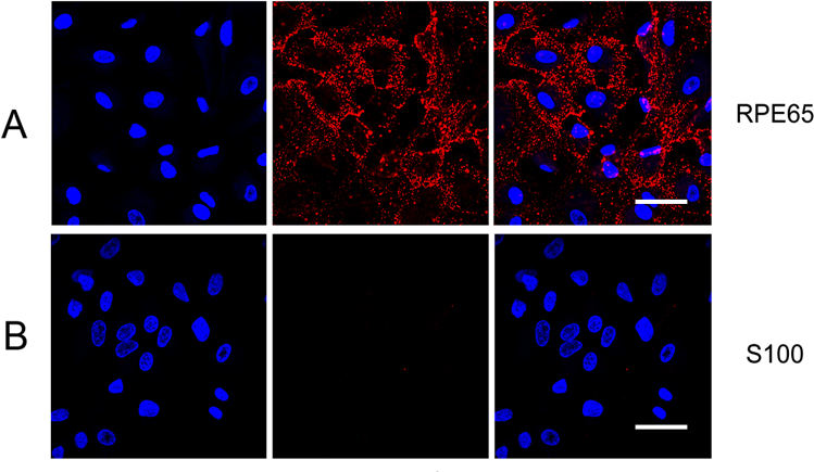
 Figure 1 of
Cheng, Mol Vis 2015; 21:939-947.
Figure 1 of
Cheng, Mol Vis 2015; 21:939-947.