Figure 1 of
Xu, Mol Vis 2015; 21:930-938.
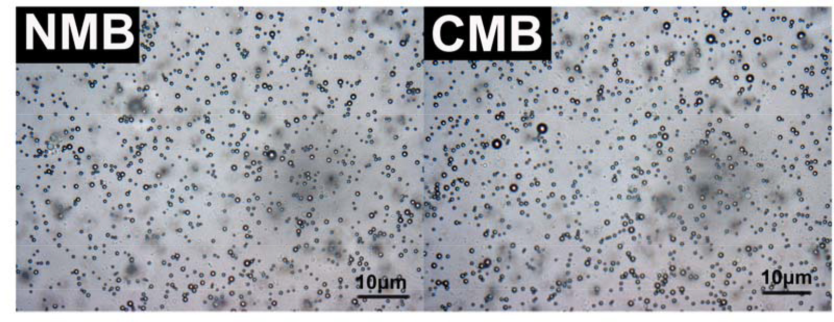
Figure 1.
Characterization of microbubbles under a microscope. Bright-field images of the neutral microbubbles (NMBs) and the cationic microbubbles (CMBs). The images were acquired at 1,000X magnification, and the scale bar is set to 10 μm.
 Figure 1 of
Xu, Mol Vis 2015; 21:930-938.
Figure 1 of
Xu, Mol Vis 2015; 21:930-938.