Figure 4. In the upper two panels, an OCT image of a 5-mm horizontal scan shows a distinct and continuous IS/OS line, correlating with
the 20/20 visual acuity in the right eye of patient IV:2 from family RPT65. In the lower two panels, intraretinal cysts in
the intermediate retina and abnormally structured IS/OS line correlate with the mother’s (II:1) poorer visual acuity in the
right eye (20/30).
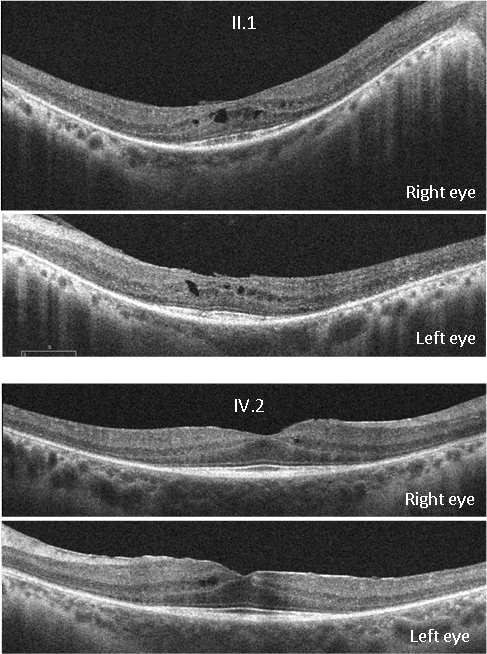
 Figure 4 of
de Sousa Dias, Mol Vis 2015; 21:857-870.
Figure 4 of
de Sousa Dias, Mol Vis 2015; 21:857-870.