Figure 4. UV-blocking spectacle lens ameliorate UVB-induced damage of ocular surface and inflammation. A-H: Histological analysis showing disordered corneal surface structure and inflammation in the unprotected cornea, but not in
the CR-39™ spectacle lens-protected cornea. The polymorphonuclear (PMN) leukocyte infiltration were found in the stromal layer
(indicated by arrow in B). Quantitative analyses of the corneal epithelium thickness in I and polymorphonuclear leukocytes infiltration in J among the study group (n = 8 per group). K-V: Immunostaining showing evident inhibition of UV-induced loss of corneal cells (P63+ basal cells and CK-5+ epithelial cells)
and UV-induced apoptosis by CR-39™ spectacle lens protection. W-Z: PAS staining showing that the UV-induced reduction of conjunctival goblet cells was significantly prevented by CR-39™ spectacle
lens protection. AA: Quantitative analyses of goblet cells within the fornix conjunctival epithelium among the study group (n = 8 per group).
EP: corneal epithelium. ST: corneal stroma. All scale bars: 25 μm.
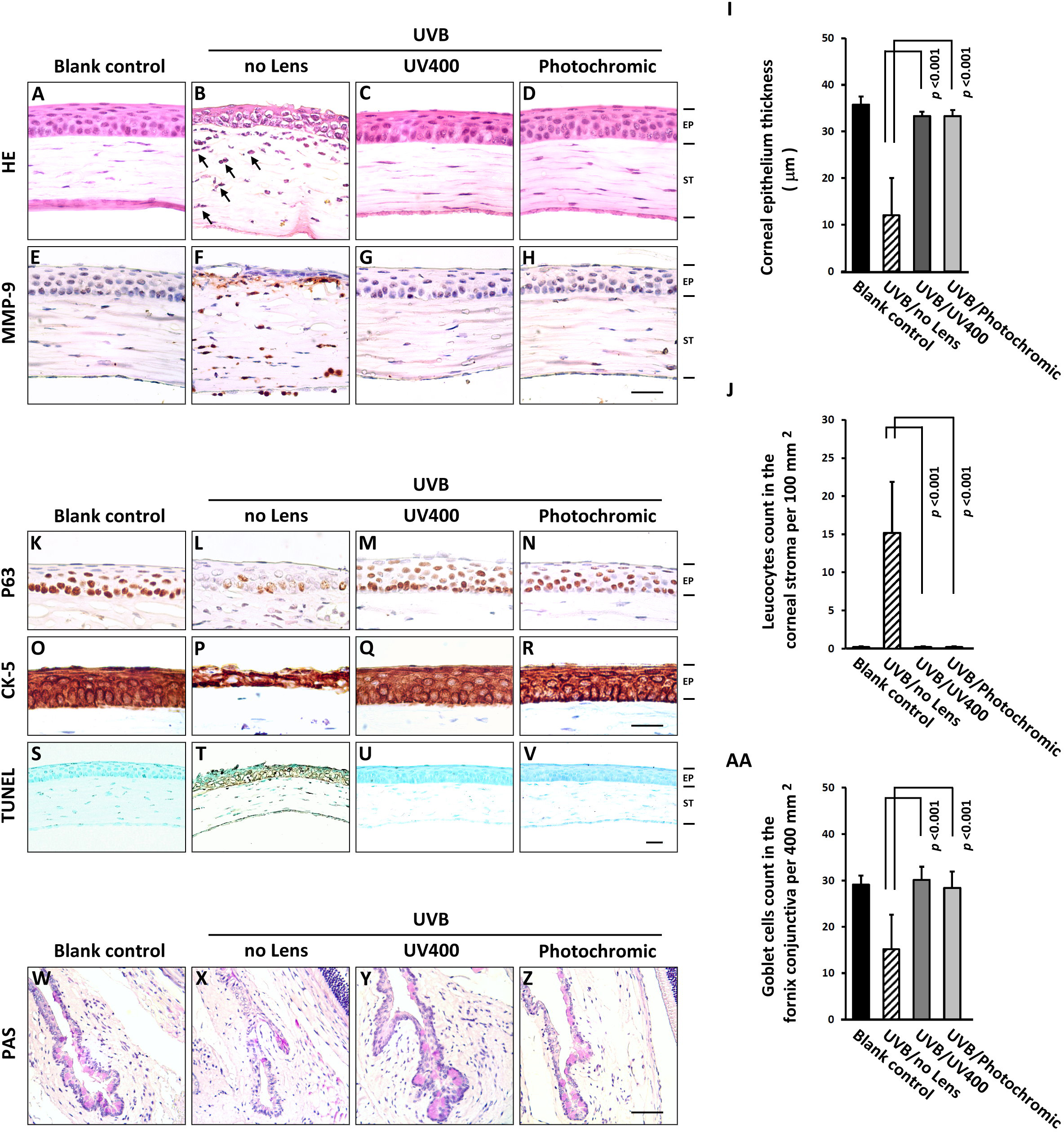
 Figure 4 of
Liou, Mol Vis 2015; 21:846-856.
Figure 4 of
Liou, Mol Vis 2015; 21:846-856.