Figure 2. Representative photos for corneal surface evaluation among the experimental groups after daily treatments of UVR (0.72J/cm2/daily) for a period of 7 days. A-D: The corneal smoothness. E-H: The corneal topography. I- L: the corneal opacity and M-P: the corneal lissamine green staining among the four study groups were assessed as parameters for the in vivo UVB protective
properties. The photos in the bottom row of M, N, O, and P are the corresponding negative images. Q, R, and S: Quantitative analyses (n = 10 per group) were also performed. The results show that all scores were reduced with the shield
protective effects from spectacle lens. All scale bars: 1.25 mm.
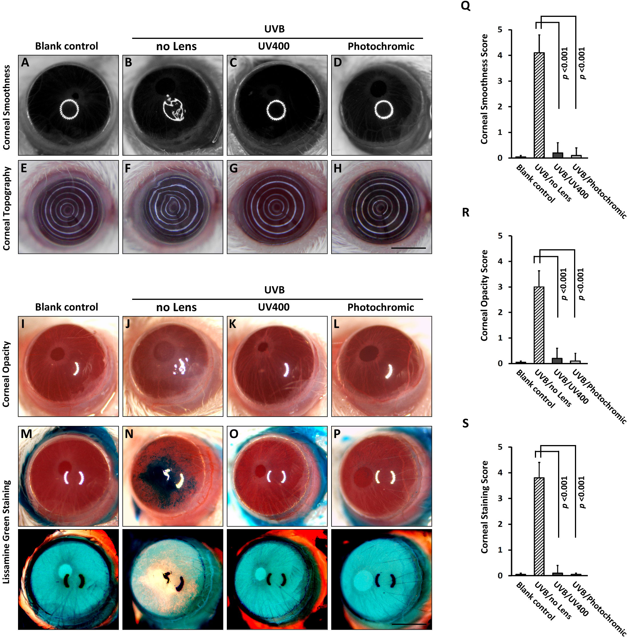
 Figure 2 of
Liou, Mol Vis 2015; 21:846-856.
Figure 2 of
Liou, Mol Vis 2015; 21:846-856.