Figure 1. Comparison of retinal morphologies of 3-week-old and 23-week-old FVB/N and C3H/HeOu mice. A–D: Longitudinal retinal cryosections stained with hematoxylin and eosin (left panels) and against S-opsin and 4',6-diamidino-2-phenylindole
(DAPI) (right panels). E–H: Retinal whole mounts stained against S-opsin. The outer nuclear layer (ONL) is two to three cell layers thick in 3-week-old
C3H/HeOu mice (A), whereas the ONL of the FVB/N mice had only one layer of remnant photoreceptors (B). In 23-week mice of both lines, the ONL was one cell layer thick (C,D). At 3 weeks of age, the OS are more ordered and cylindrical in the C3H/HeOu retinas (A,E) compared to the FVB/N retinas (B,F). At 23 weeks (G,H), the OS are lost, and S-opsin is located in the cell bodies. Note that the FVB/N retina contains markedly fewer cones. Scale
bars, (A–D) 20 μm, (E–H) 50 μm.
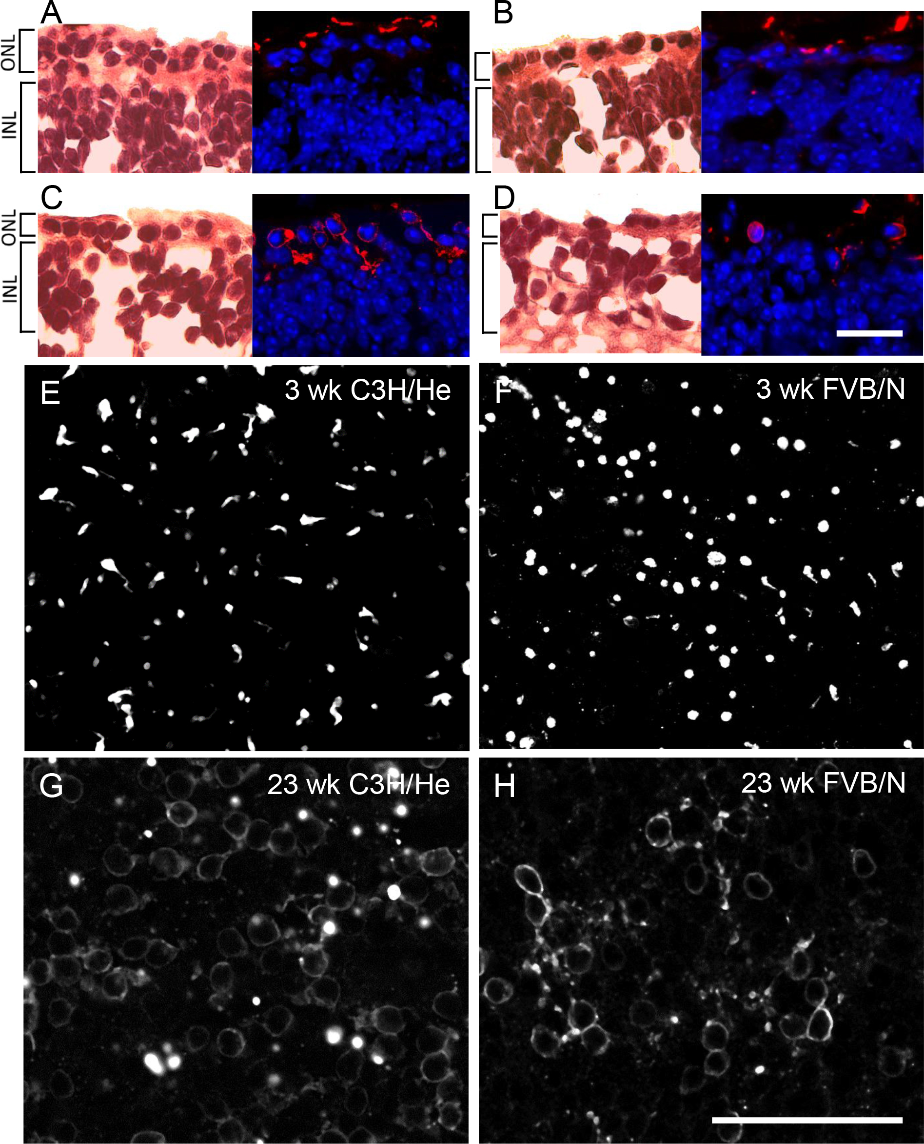
 Figure 1 of
van Wyk, Mol Vis 2015; 21:811-827.
Figure 1 of
van Wyk, Mol Vis 2015; 21:811-827.