Figure 8. Ultrastructure of the posterior corneal stroma of WT and mutant embryos at E18.5. A: Lamellar structure of piled elongated keratocytes and collagenous connective tissue are observed in the posterior stroma
of the wild-type (WT) embryo cornea. B: This lamellar structure is not observed in the anterior stroma of the mutant cornea (asterisk). Neovascularization (NV)
is also seen in the mutant stroma. Bar, 5 μm.
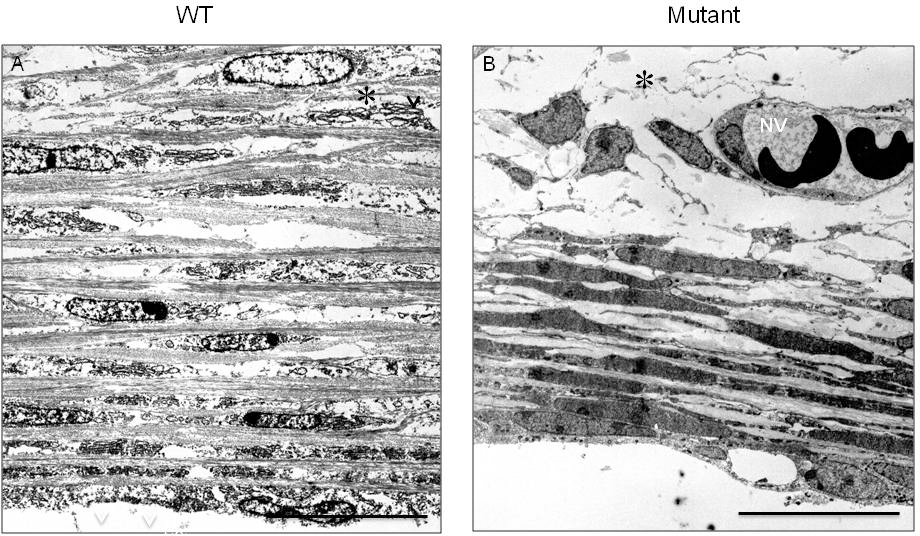
 Figure 8 of
Mizoguchi, Mol Vis 2015; 21:793-803.
Figure 8 of
Mizoguchi, Mol Vis 2015; 21:793-803.