Figure 5. Immunohistochemistry for laminin in the E18.5 corneal epithelium and its ultrastructural histology. Immunohistochemistry detects
laminin in the epithelial basement membrane of the corneal epithelium (arrowheads) in the E18.5 wild-type (WT) embryo (A), while the epithelial nodules that grows downward to the stroma lacks a laminin-basement membrane (arrows; B). Ultrastructural observation shows that the corneal epithelium (C, Epi) forms stratification of the intraepithelial-differentiated cell in the WT tissue, while it consists of disarranged
spheroidal epithelial epidermal cells (D, Epi) in the mutant tissue. Mutant epithelial cells in the mutant mouse lack upward differentiation. The epithelial–stromal
interface is irregular in the mutant tissue with disorganized stromal connective tissue (Stroma) in the mutant cornea. Higher
magnification observation shows that the WT corneal epithelium (Epi) has a basement membrane (arrows) between stroma (E), while the basal epithelial cells (Epi) lacks this structure at the interface with the underlying stroma in the mutant (F). Bar, 50 μm (A, B); 5 μm (C, D); 500 nm (E, F).
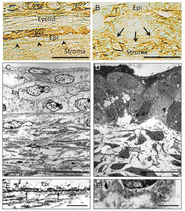
 Figure 5 of
Mizoguchi, Mol Vis 2015; 21:793-803.
Figure 5 of
Mizoguchi, Mol Vis 2015; 21:793-803.