Figure 4. Effect augmented β-catenin on histology of the cornea epithelium. a. Hematoxylin and eosin (HE) staining histology suggested
that at E13.5 there is no obvious difference of the corneal epithelial morphology between a wild-type (WT) littermate and
the mutant of gof-β-catenin in epithelial tissue (A, B). At E15.5, the WT corneal epithelium is thin (C), while the epithelium of the mutant mouse embryo seemed thicker than that in the WT control, and localized multilayerization
was observed (D). At E17.5 and E18.5, focal epithelial cell nodules (arrows) are observed in the corneal epithelium in the mutant embryo
(F,H), and such structures are not seen in the WT tissue (E, G). Throughout the embryonic day points, cell density in the corneal stroma seems higher than that of the WT stroma. Bar, 50
μm (A, B); 100 μm (C–H). Expression of β-catenin is not seen in the corneal epithelium of the WT embryo (I) at E13.5. Obvious β-catenin immunoreactivity is observed in the mutant corneal epithelium (arrows) at this day point (J). At E15.5, β-catenin is detected in the WT corneal epithelium (K). Marked β-catenin staining is detected in the thickened epithelium and more markedly in the focal cellular nodules (arrows)
in the epithelium of the mutant embryo (L). At E17.5 and E18.5, the expression of β-catenin is faint in the WT corneal epithelium (M, O), while the mutant epithelium exhibits β-catenin immunoreactivity in the proliferative nodules (arrows), but not the thickened
epithelium among each nodule (N, P). The insert in Frame F indicates that β-catenin immunoreactivity is detected in the cytoplasm and nuclei (arrows) of the
epithelial cells in the nodular structure. Epi, corneal epithelium; Bar, 50 μm (A, B, I, J); 100 μm (C–H, K-P).
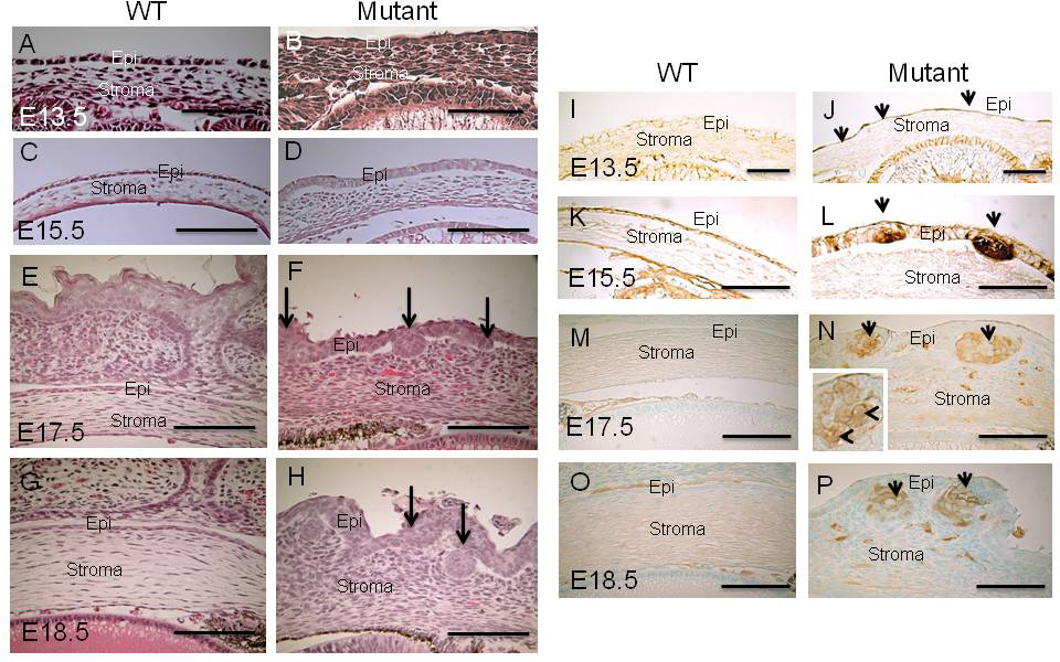
 Figure 4 of
Mizoguchi, Mol Vis 2015; 21:793-803.
Figure 4 of
Mizoguchi, Mol Vis 2015; 21:793-803.