Figure 3. Ultrastructural and immunohistochemical examinations of impaired eyelids in mutant mice. Hematoxylin and eosin histology shows
a clear difference in the cellular architecture of the epidermis and the mesenchyme of the eyelid (anlage) between the wild-type
(WT) embryo (A) and the mutant embryo (B) with gain-of-function (gof)-β-catenin at E18.5. Nodular structures (srar) are observed in the mutant epidermis, but not
in the WT epidermis. Mesenchymal cells are more densely packed (asterisk) in the mutant eyelid anlage than in the WT eyelid.
Ultrastructural observation showed loss of stratification or intraepidermal differentiation in the upper layers with relatively
round basal-like cells (Epi) and reduction of the accumulation of the collagenous matrix in the dermal tissue (asterisks)
in an E18.5 mutant embryo (D) compared with the WT tissue (C). Immunohistochemistry shows laminin is well condensed in the subepithelial basement membrane zone in the WT embryo (E), while immunoreactivity for laminin is disrupted (arrows) beneath the epithelial nodular structure in the mutant eyelid
anlage (F). The accumulation of β-catenin is detected in the nodular structures of the epidermis of the mutant embryo at E18.5 (arrows,
H), while faint staining for β-catenin was seen in the epidermis of the WT embryo (G). The eyelid epidermis in the E18.5 WT and mutant embryos is not labeled for vimentin (I, J). The population of bromo-deoxyuridine (BrdU)-labeled epidermal keratinocytes was similar in the eyelid epidermis of the
WT (K) and mutant embryos (L) at E15.5. Bar, 5 μm (C, D); 20 μm (E, F); 50 μm (A, B, G–J); 100 μm (K, L).
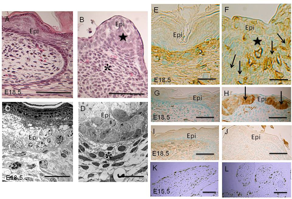
 Figure 3 of
Mizoguchi, Mol Vis 2015; 21:793-803.
Figure 3 of
Mizoguchi, Mol Vis 2015; 21:793-803.