Figure 2. HE histology of eyes of a WT embryo and a mutant embryo with gof-β-catenin in epithelial tissues. Hematoxylin and eosin (HE)
histology shows a clear difference in the cellular architecture of the eyelid anlage and the cornea between the wild-type
(WT) embryo and the mutant embryo with gain-of-function (gof)-β-catenin. No obvious difference in the structure of the eye
and eyelids between the WT (A) and mutant embryos (B) was observed at E13.5. At E15.5, the eyelid (arrows) development is impaired (D) compared with the WT tissue (C). At E17.5 and E18.5, the cornea is completely covered with fused eyelids (arrows) in the WT embryo (E, G), while no eyelids, but just anlage (arrow) of an eyelid, are observed in the mutant (F, H). The surface of the cornea is thicker with an irregular surface in the mutant eye while smooth curvature is seen in the
WT embryo (compare H to G). Bar, 100 μm.
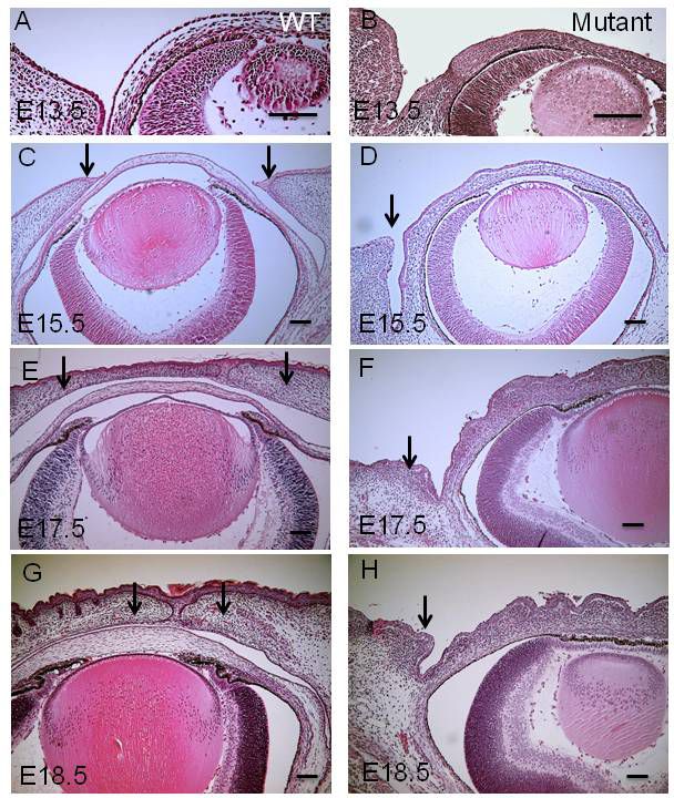
 Figure 2 of
Mizoguchi, Mol Vis 2015; 21:793-803.
Figure 2 of
Mizoguchi, Mol Vis 2015; 21:793-803.