Figure 2. AF images, color photographs, red-free photographs, and OCT examinations from subjects 10, 11, and 12. In subject 10 (A–E), the AF image (A) is normal. Fundus photographs (B and C) show subtle pigmentary changes in the macula. The OCT B-scan (D) reveals subtle central outer retinal disruption with some central thickening (E). Subject 11 has normal FA image (F) and fundus photographs (G and H). In subject 12 (I–M), the AF image (I) is normal while the fundus photographs (J and K) show pigmentary changes in the macula, and the OCT B-scan reveals some degree of central outer retinal disruption (L) and central retinal attenuation (M).
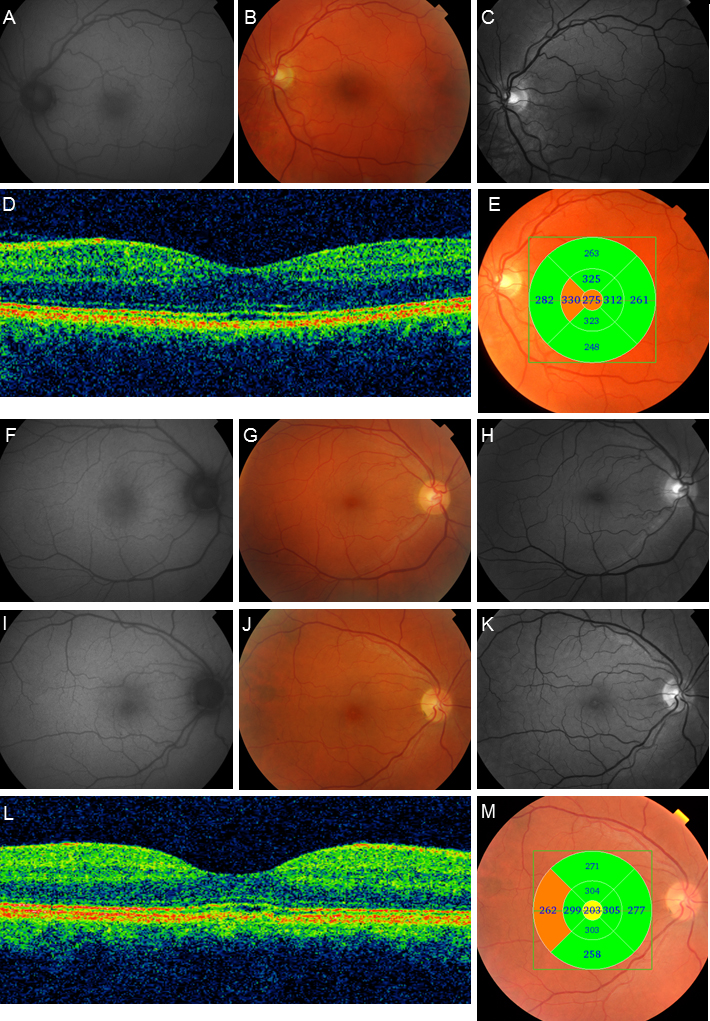
 Figure 2 of
Kjellström, Mol Vis 2015; 21:767-782.
Figure 2 of
Kjellström, Mol Vis 2015; 21:767-782.