Figure 1. Basal VEGF-A and PlGF secretion. A: In the RPE/choroid, vascular endothelial growth factor (VEGF)-A is constitutively secreted with significantly higher secretion
on the basal side. B: Placental growth factor (PlGF) is also constitutively secreted with only minor secretion on the apical and significantly
stronger secretion on the basal side. C: In the RPE cell culture, VEGF is basally and apically secreted, with a stronger secretion on the basal side. D: In RPE cell culture, no PlGF was found. E: The secretion of VEGF in the RPE cell culture is stable over time. Supernatants were collected for 24 h and were analyzed
in enzyme-linked immunosorbent assay (ELISA). E: Supernatant was collected for 24 h, and the medium was changed and recollected after another 24 h. Significance was determined
with the Student t test; +++ p<0.001. n≥30 (A–C), n=7 (D), n=10 (E, apical), n=6 (E, basal). Bars depict the mean and the standard deviation.
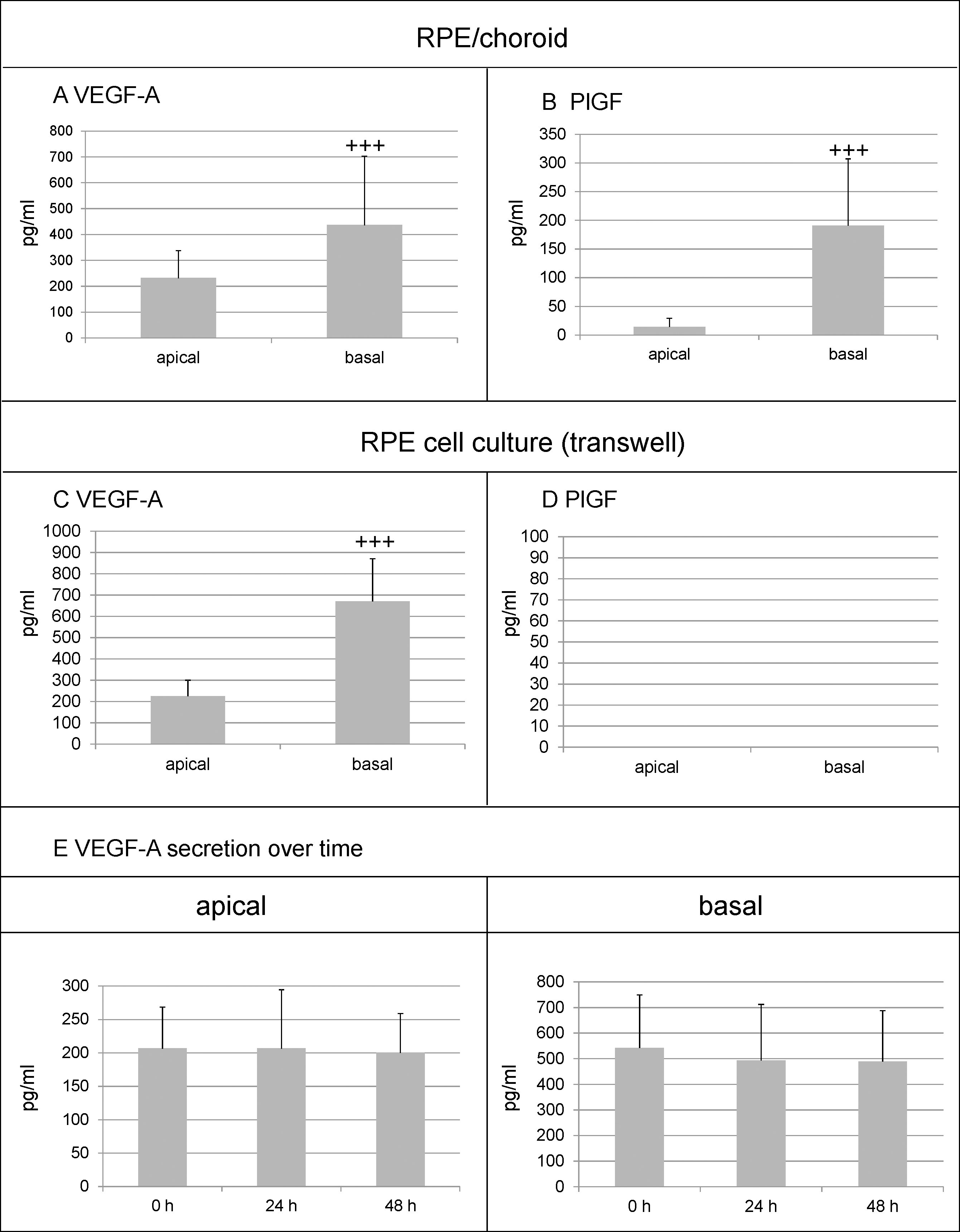
 Figure 1 of
Klettner, Mol Vis 2015; 21:736-748.
Figure 1 of
Klettner, Mol Vis 2015; 21:736-748.