Figure 1. WWOX expression in primary and recurrent pterygium. Immunohistochemical staining of WWOX expression in the head and body of
primary and recurrent pterygium and the conjunctiva (n=8/group). A: WOX1 and B: p-WOX1 are highly expressed in the epithelial cells of the pterygium head, whereas they were less expressed in the body
and the conjunctiva. WWOX expression is stronger in recurrent pterygia than in primary pterygia. Magnification: 400X. Bar=25
μm.
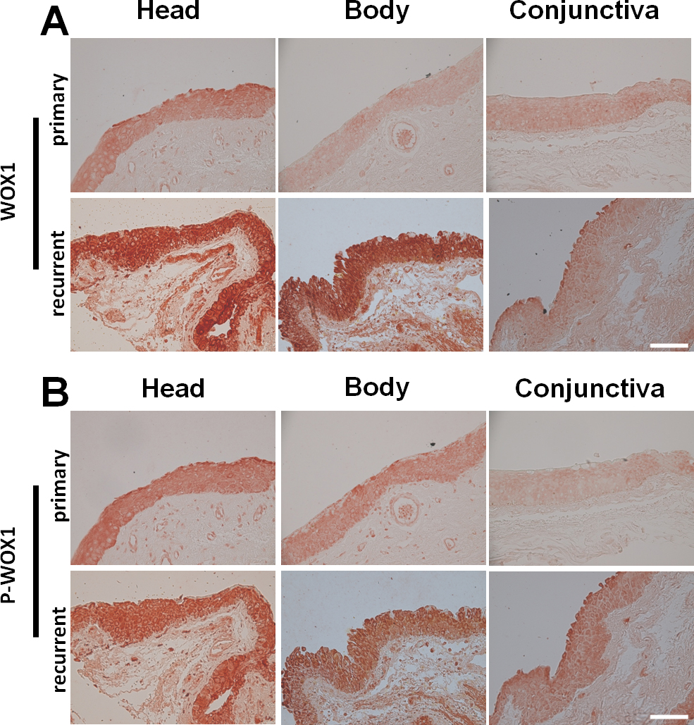
 Figure 1 of
Huang, Mol Vis 2015; 21:711-717.
Figure 1 of
Huang, Mol Vis 2015; 21:711-717.