Figure 5. Isolation of CD31-positive cells from C-FVMs. Immunofluorescence localization of isolectin (A) and CD31 (B) cells from clinical case 4 identifies rare positive cells. Fibrovascular membrane (FVM) from clinical case 5 processed for
immunohistochemistry using antibodies against CD31 (red) and counterstained with hematoxylin (blue; C). Light micrographs of C-FVMs from clinical case 5 show isolation of CD31-positive cells, bound to CD31-antibody coated Dynabeads
before cell culture (D). Scale bar=50 µm.
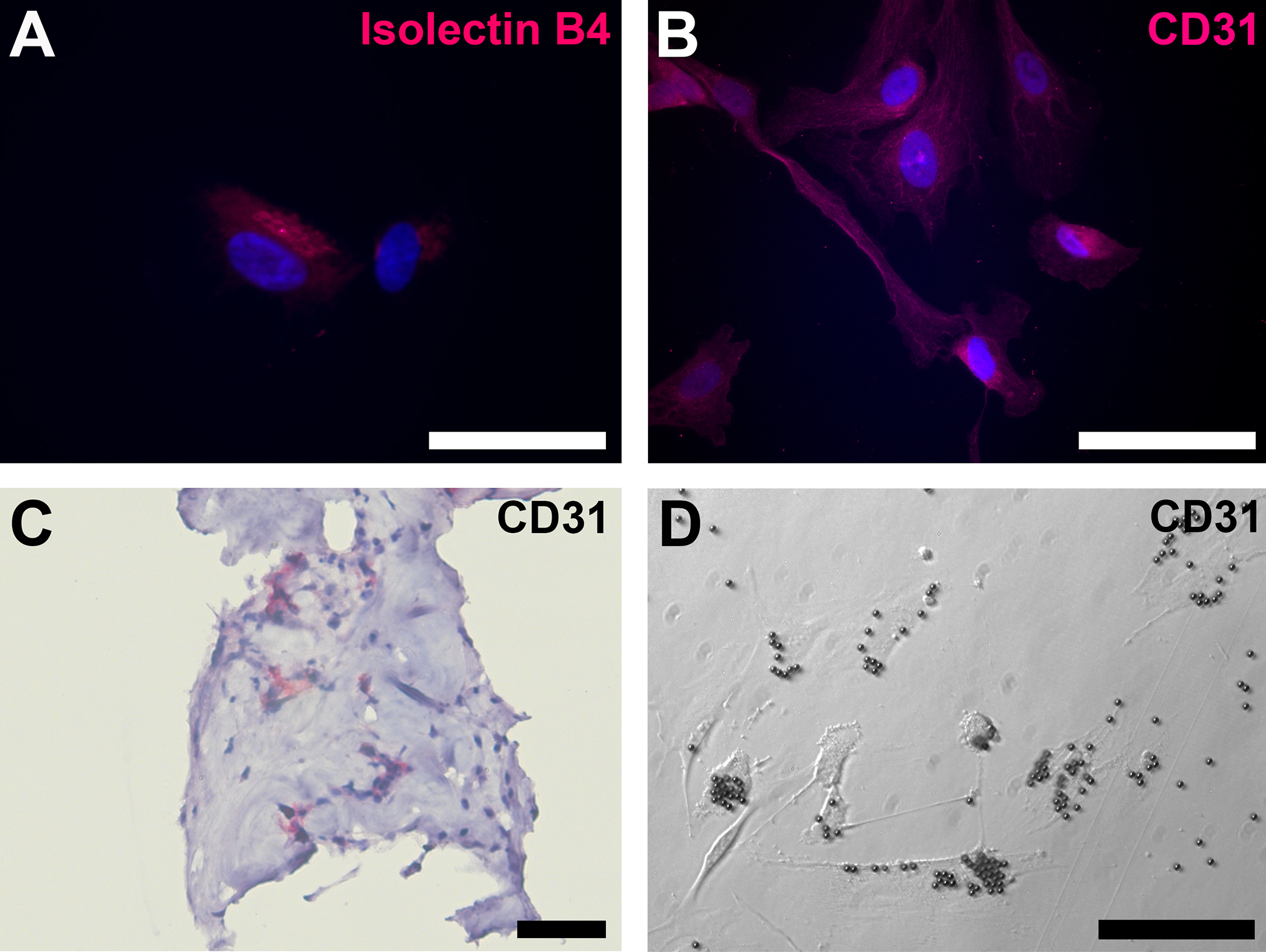
 Figure 5 of
Kim, Mol Vis 2015; 21:673-687.
Figure 5 of
Kim, Mol Vis 2015; 21:673-687.