Figure 4. Proliferative activity of C-FVMs varies between different clinical cases. Fibrovascular membranes (FVMs) from clinical cases
3 (A) and 5 (B) were processed for immunohistochemistry using antibodies against Ki67 (in red) and counterstained with hematoxylin (blue). C-FVMs from clinical case 3 and clinical case 5 differ in the percentage of cells that are Ki67-negative (66.06% versus
81.60%) and Ki67-positive (33.94% versus 18.40%) when cultured in growth media (C, D). The percentage of cells that are Ki67-negative (92.57% versus 94.06%) and Ki67-positive (7.43% versus 5.94%) in C-FVMs
from clinical cases 3 and 5 are similar under growth factor-starvation conditions (E, F). Scale bar=50 µm.
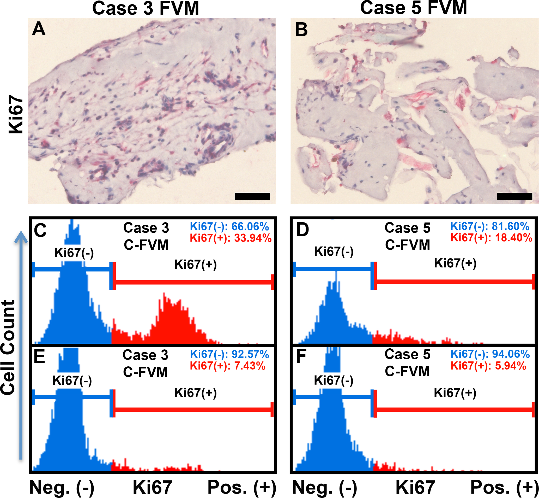
 Figure 4 of
Kim, Mol Vis 2015; 21:673-687.
Figure 4 of
Kim, Mol Vis 2015; 21:673-687.