Figure 1. Clinical case 3 representing combined tractional and rhegmatogenous retinal detachment with resection of FVM and growth of
C-FVM. Pre-operative fundus photograph of the left eye reveals combined tractional and rhegmatogenous retinal detachment of
the left eye with 20/200 vision (A). FVM (outlined by dotted white line) involves the macula and reveals fibrous tissue with aberrant blood vessels. Post-operative
fundus photograph five months post vitrectomy and one month after cataract surgery demonstrates re-attached retina with 20/25
vision (B). Bright field microscopy of surgically resected FVM shows fibrous tissues and blood vessels (C, inset). Bright field microscopy of cells derived from FVM reveals a morphologically heterogeneous mixture of fibroblastic
and stellate cells (D). Scale bar = 100 µm.
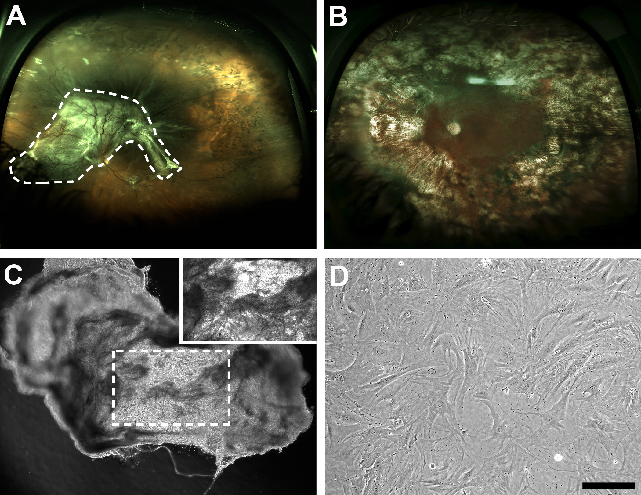
 Figure 1 of
Kim, Mol Vis 2015; 21:673-687.
Figure 1 of
Kim, Mol Vis 2015; 21:673-687.