Figure 1. The box-and-whisker plot represents the median and minimum to maximum range of vitreous LRP6 or VEGF levels. Vitreous samples
were measured among 30 subjects with PDR and 25 subjects as control. The levels of (A) lipoprotein receptor-related protein 6 (LRP6) and (B) vascular endothelial growth factor (VEGF) were significantly increased in eyes with proliferative diabetic retinopathy (PDR)
compared to controls (LRP6: p=0.002 and VEGF: p<0.001, respectively).
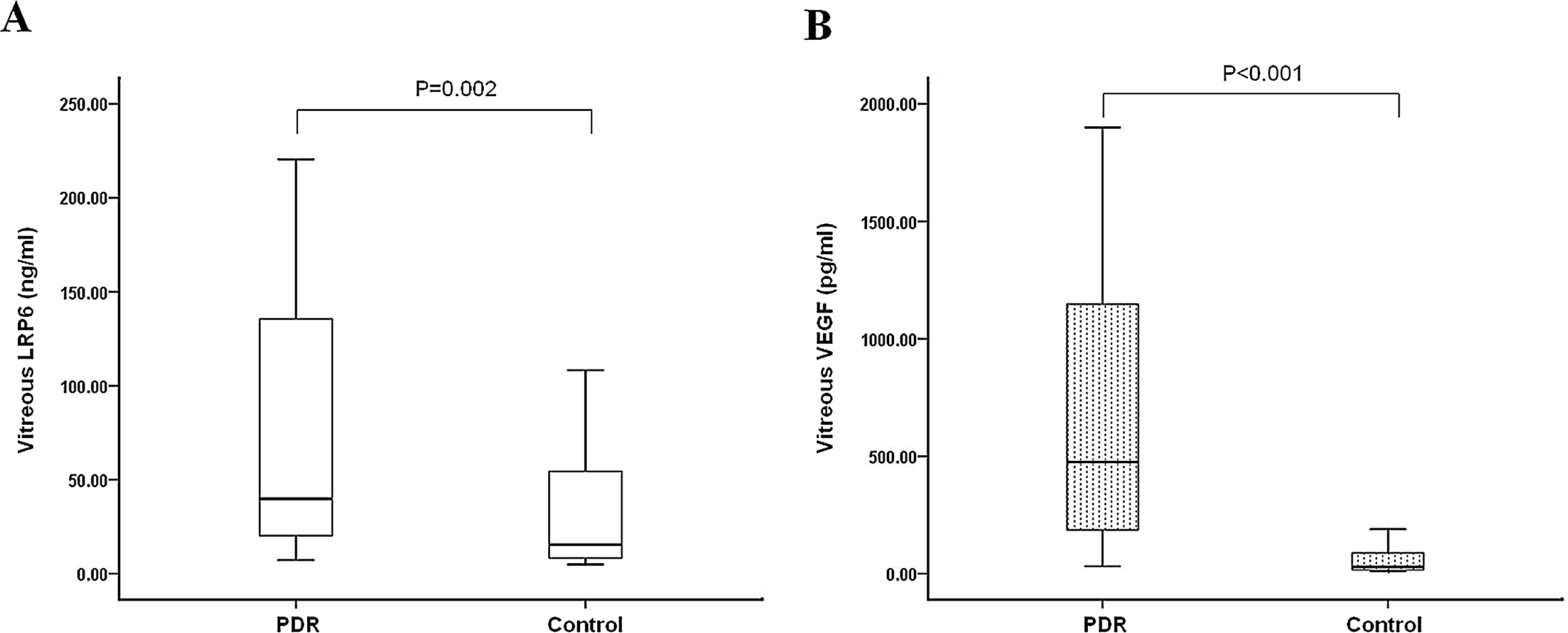
 Figure 1 of
Gao, Mol Vis 2015; 21:665-672.
Figure 1 of
Gao, Mol Vis 2015; 21:665-672.