Figure 3. p63 and ABCG2 positivity in both healthy and pathological condition. A: Nuclear p63 staining in healthy tissues was shown. B: p63 expression was evidenced also in pathological cases. C: Cytoplasmic ABCG2 positivity was observed healthy samples. D: ABCG2 expression was achieved also in inflamed tisses. Arrowheads indicate inflammatory cells. Each section was counterstained
with hematoxylin. Bar scale=50 micron. Magnification X200. * Indicates the limbal side of the specimen.
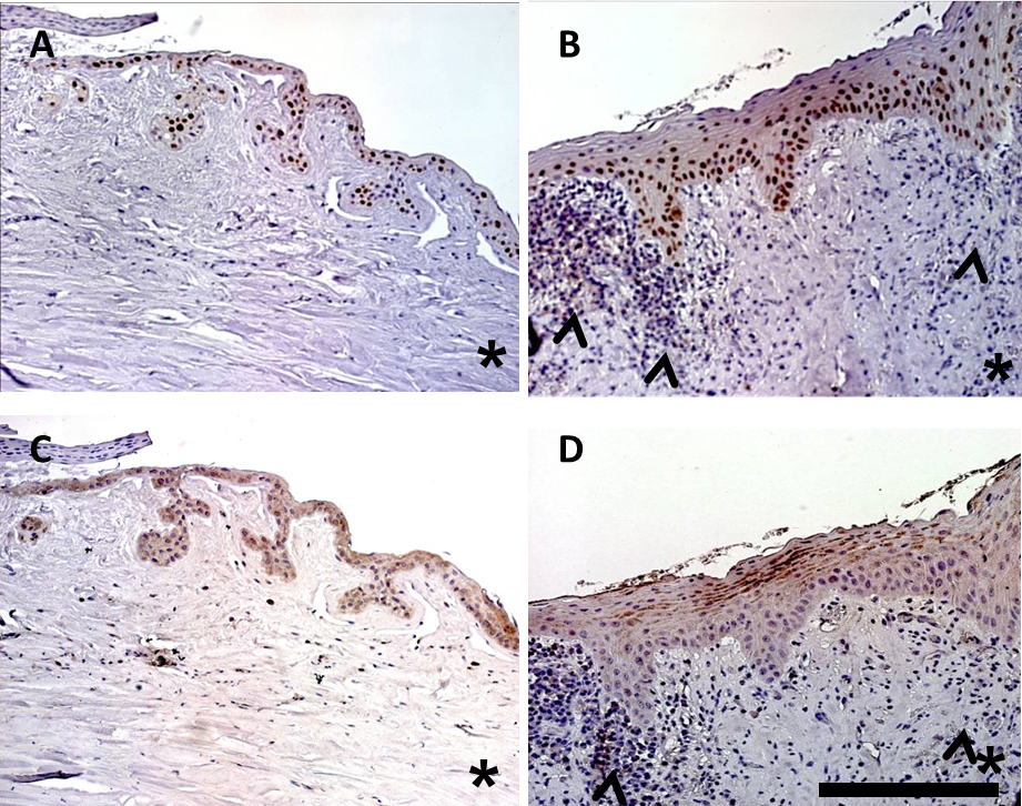
 Figure 3 of
Curcio, Mol Vis 2015; 21:644-648.
Figure 3 of
Curcio, Mol Vis 2015; 21:644-648.