Figure 3. ZO-1, ZONAB, and F-actin expression in collagenase A-prepared BCECs with or without Y-27632 on days 1, 3 (subconfluence),
and 7 (confluence). In the collagenase A-prepared bovine corneal endothelial cells (BCECs) without Y-27632, the TJ protein
ZO-1 developed gradually from the nucleus to the cell–cell contact and formed a continuous hexagonal pattern, as it did in
the TrypLE-prepared BCECs without Y-27632. The ZONAB expression exhibited staining in the nucleus that was stronger that in
the TrypLE-prepared BCECs in the beginning, as in the ZO-1 expression at the cellular borders (A, B: day 1; D, E: day 3; G, H: day 7). The F-actin cytoskeleton was arranged into a dense peripheral band at the cell margin and cortical actin mat with
prominent perinuclear staining in the beginning (C, F, I). The effect of Y-27632 on the expression of the ZO-1 (A’, D’, G’), ZONAB (B’, E’, H’), and F-actin cytoskeleton (C’, F’, I’) was similar to that without Y-27632. Similarly, the cellular morphology became irregular (G, G’), and there was a marked decrease in the amount of F-actin in the presence of Y-27632 compared with the BCECs without Y-27632
in the beginning (I, I’). The ZONAB expression patterns were similar in the BCECs with and without Y-27632 (B, E, H, B’, E’, H’).
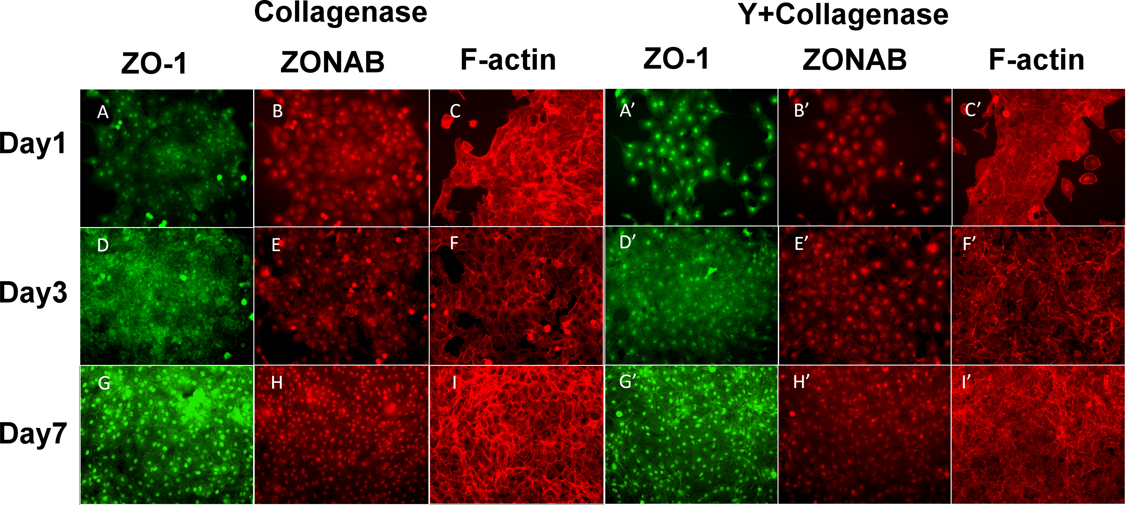
 Figure 3 of
Su, Mol Vis 2015; 21:633-643.
Figure 3 of
Su, Mol Vis 2015; 21:633-643.