Figure 2. Multiphoton images of the mouse retina, with the THG signal shown in red and the TPAF signal shown in green. A: A sagittal/transverse retinal section of an albino (BALB/c) mouse eye, showing simultaneous third harmonic generation (THG)
and two-photon autofluorescence (TPAF) signals from different layers of the retina. A significant THG signal is detected in
the RPE layer, outer nuclear layer (ONL), inner nuclear layer (INL), and ganglion cell layer (GCL). The TPAF signal is predominantly
generated at the photoreceptors (PRs), the outer plexiform layer (OPL), and the inner plexiform layer (IPL). B: The outer segment (OS), inner segment (IS), and cell nuclei (*) of the photoreceptors are resolved with THG (the RPE layer
detached during processing). C: Individual cellular nuclear membrane (x) of the INL resolved with THG. D: Individual cellular nuclear membrane (x) of the ganglion cells resolved with THG. E: A sagittal/transverse retina section from a pigmented (C56BL/6) mouse eye. A strong THG signal originates from the pigment
granules (+) within the RPE of the pigmented mouse sections, a signal not seen from the RPE layer within the albino sections
in Panels A and B. Post-processing filters were applied for better visualization of the TPAF signal, which we believe are the photoreceptor
cytoplasmic material or axons. Scale bar=10 µm.
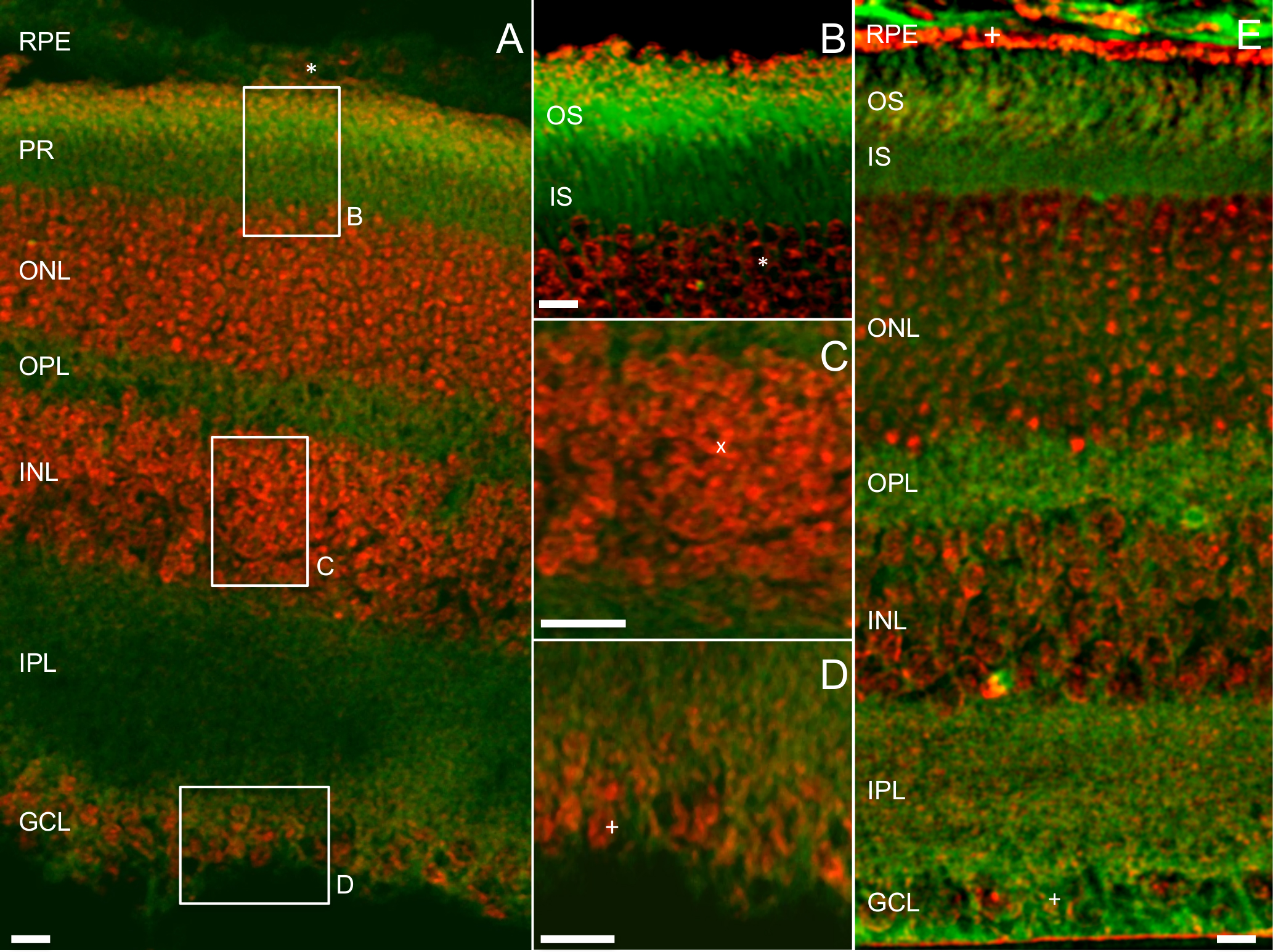
 Figure 2 of
Masihzadeh, Mol Vis 2015; 21:538-547.
Figure 2 of
Masihzadeh, Mol Vis 2015; 21:538-547.