Figure 1. Multiphoton microscopy setup for simultaneous third harmonic generation (THG) and two-photon autofluorescence (TPAF) imaging
of retina. A: A femtosecond fiber laser oscillator generates a 1044-nm laser beam to target the cross sections of full-thickness mouse
retina. The laser beam is scanned and focused across the sample with a 60X microscope objective (OBJ). A dichroic mirror (DM1)
separates the excitation laser beam (1044 nm) from the TPAF signal (522 to 600 nm) traveling in the backward (epi) direction
and detected with a photomultiplier (PMT1). A high numerical aperture condenser lens (CL) collects the forward propagating THG signal. A second dichroic mirror and
a narrow band filter sets separate the forward propagating TPAF and excitation beams from the THG signal (348 nm), which is
detected with a photomultiplier (PMT2), DM1: dichroic mirror, SL: scanning lens, TL: tube lens, OBJ: objective, S: sample, CL: condenser lens, DM2: dichroic and
narrow band filter, PMT: photomultiplier. B: A Jablonski diagram showing the interaction of multiple infrared photons between the electronic ground state and the electronic
virtual state. In THG, three infrared excitation photons are instantaneously up-converted into a single photon of three times
the energy. C: Under multiphoton microscopic conditions, no THG signal is generated inside a homogenous medium, i.e., the aqueous media
of the cytoplasm. D: At the interface of two media with different nonlinear susceptibilities, i.e., the aqueous media and the lipidic organelle,
significant THG is generated at the focus of the excitation beam. E: The THG signal from the lipid-rich nuclear membrane (+), cell membrane (*), and lipid droplets (x) of an opossum kidney
(OK) cell create a biomarker for lipid-specific functional microscopy. F: THG signal from the pigment granules (x) of a cultured human fetal RPE (hfRPE) cell. The region with no THG signal (+) represents
the nuclei.
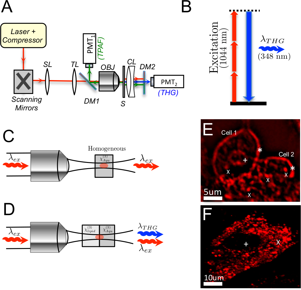
 Figure 1 of
Masihzadeh, Mol Vis 2015; 21:538-547.
Figure 1 of
Masihzadeh, Mol Vis 2015; 21:538-547.