Figure 5. Phosphorylation of Erk and Akt induced by TCS-401. A, B: RPE cells were incubated with 0.5, 1, and 2 μM TCS-401 for 30 min for an assay of Erk and Akt phosphorylation, and levels
of phosphorylated and total Erk and Akt were determined with western blot analysis, respectively. C, D: RPE cells were pretreated with 20 μM PD98059 for 30 min and then incubated with 2 μM TCS-401 for 30 min for an assay of
Erk phosphorylation using western blot. E, F: RPE cells were pretreated with 10 μM LY294002 for 30 min and then incubated with 2 μM TCS-401 for 30 min for an assay of
Akt phosphorylation using western blot. The data represent the mean±standard deviation of three independent experiments. *,
#p<0.05, compared to the control group; &p<0.05, compared to treatment with only TCS-401. Akt, protein kinase B; Erk, extracellular signal-regulated kinase; RPE, retinal
pigment epithelium.
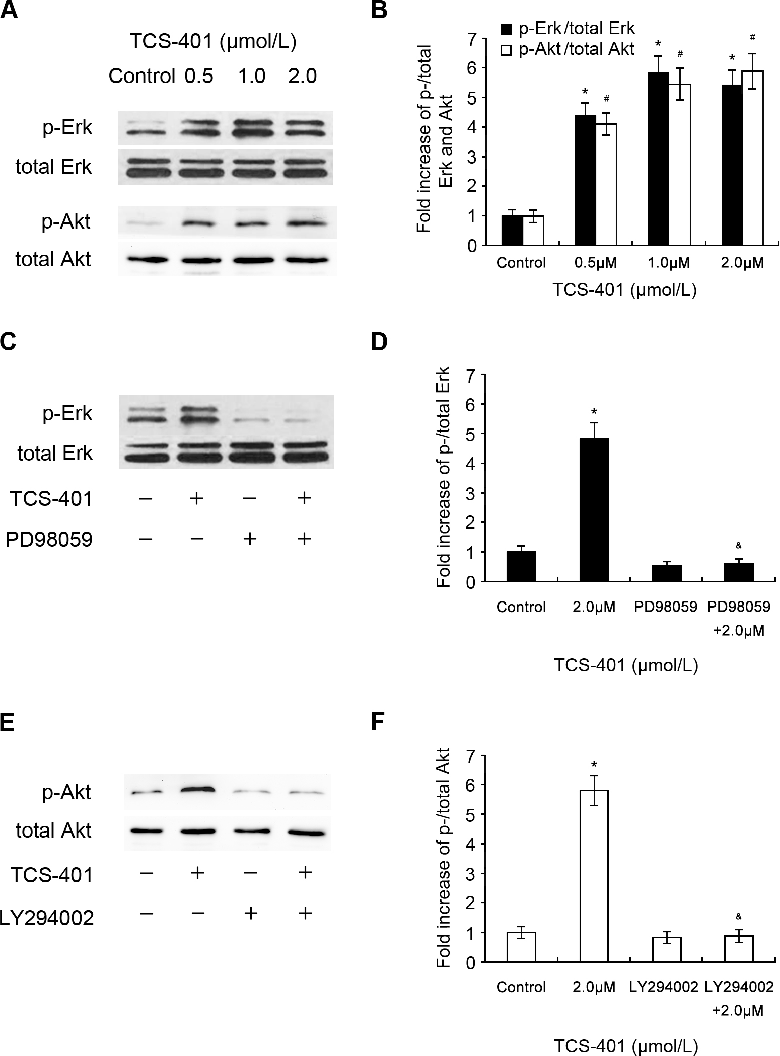
 Figure 5 of
Du, Mol Vis 2015; 21:523-531.
Figure 5 of
Du, Mol Vis 2015; 21:523-531.