Figure 1. Immunolocalization of PTP1B in RPE cells of a rat model of retinal detachment. A: Fundus photograph shows half-side retinal detachment (arrow) without retinal breaks. B, C: Immunolocalization of PTP1B in stretched preparations of rat RPE (B: non-retinal detachment area, C: retinal detachment area). The data are representative of at least three independent experiments. Original magnification=400X.
PTP1B, protein tyrosine phosphatase 1B; RPE, retinal pigment epithelium.
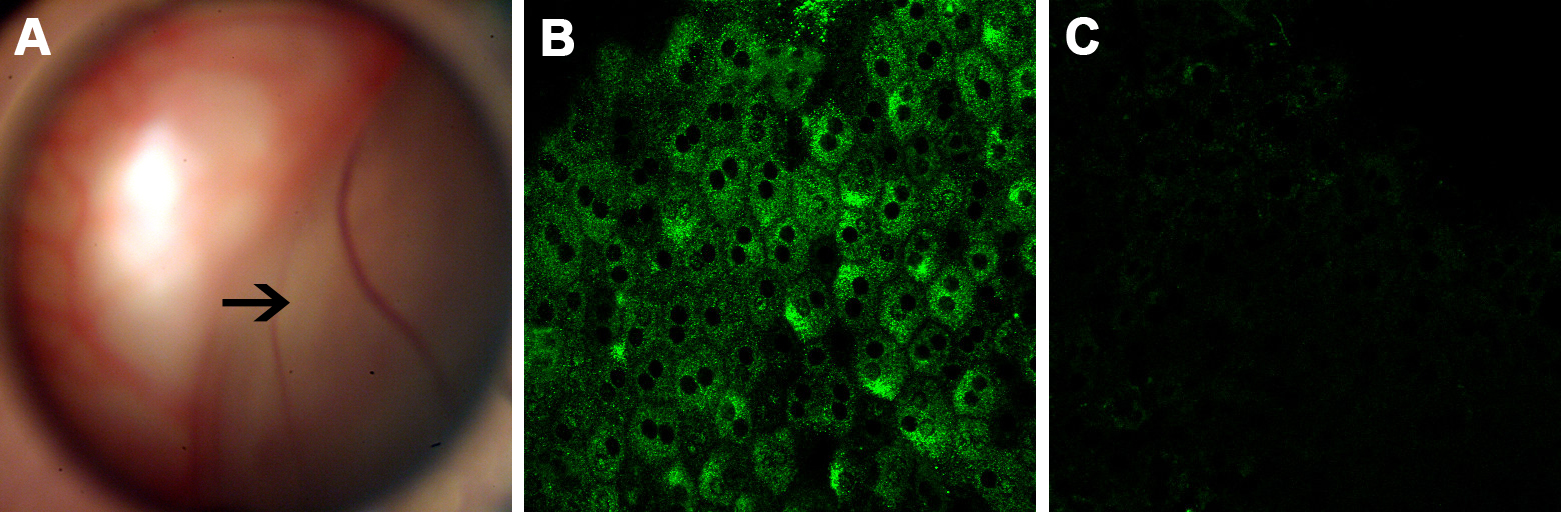
 Figure 1 of
Du, Mol Vis 2015; 21:523-531.
Figure 1 of
Du, Mol Vis 2015; 21:523-531.