Figure 2. Longitudinal imaging of a TAg-RB tumor with OCT. A single, representative eye imaged weekly from 2 to 12 weeks of age as indicated
shows early clusters of presumptive T-antigen (TAg)-positive cells decreasing around 4 weeks, as previously observed with
immunohistochemistry. Then, a distinct tumor mass is first evident at 5 weeks and grows over subsequent weeks. Retinal blood
vessels (seen by their shadow) sit adjacent to this small tumor during weeks 6–7 and atop the tumor from week 8. Optical coherence
tomography (OCT) scale bars=100 µm.
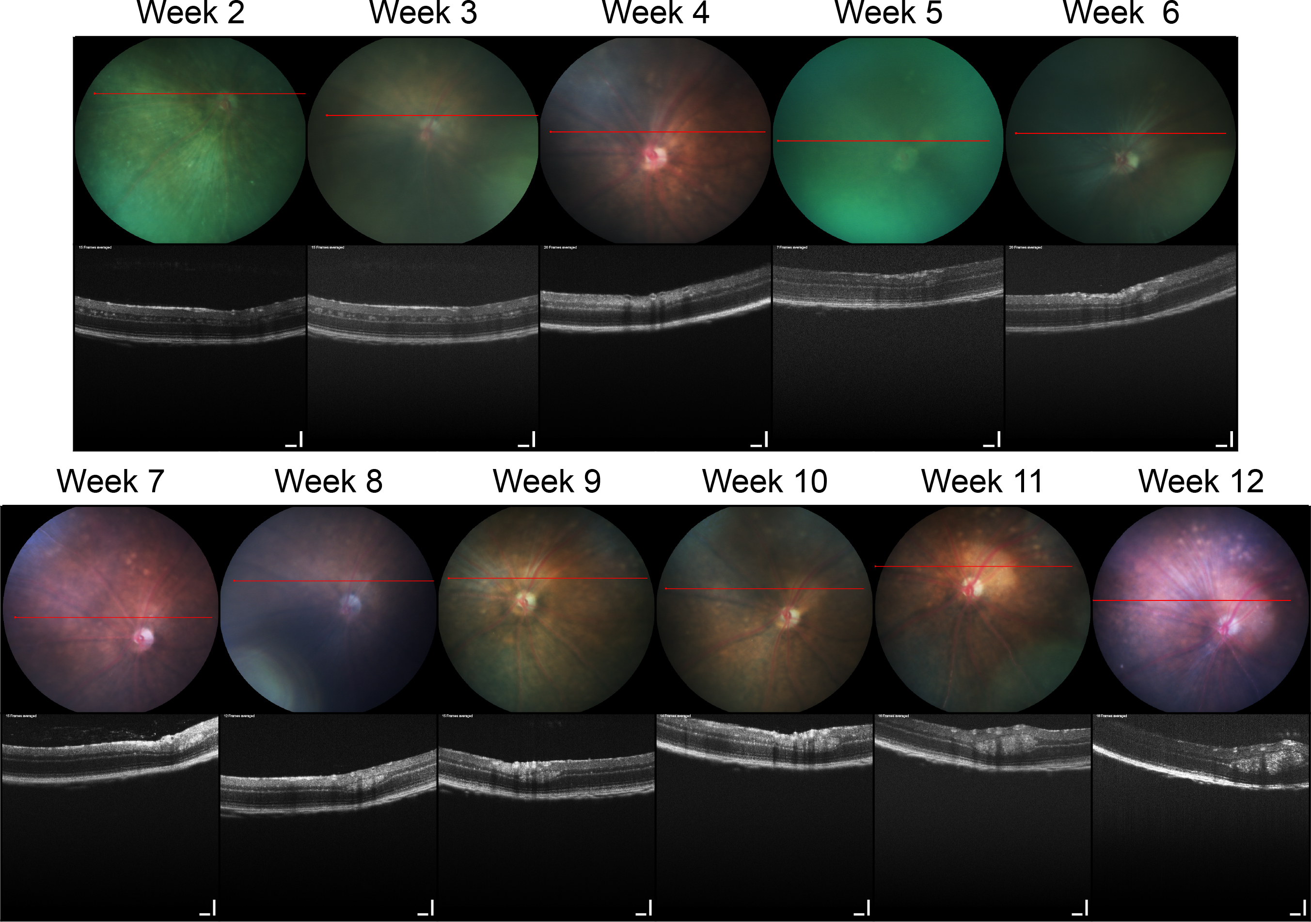
 Figure 2 of
Wenzel, Mol Vis 2015; 21:515-522.
Figure 2 of
Wenzel, Mol Vis 2015; 21:515-522.