Figure 1. Earliest areas of T-antigen positive cells evident in TAg-RB retinas with OCT. At 2 (A, B) and 3 (D, E) weeks of age, hyperreflective clusters are seen in the inner nuclear layer of T-antigen retinoblastoma (TAg-RB) but not
(C, F) wild-type eyes with optical coherence tomography (OCT) (bottom). All fundi appear normal at this age (top). OCT scale bars=100
µm.
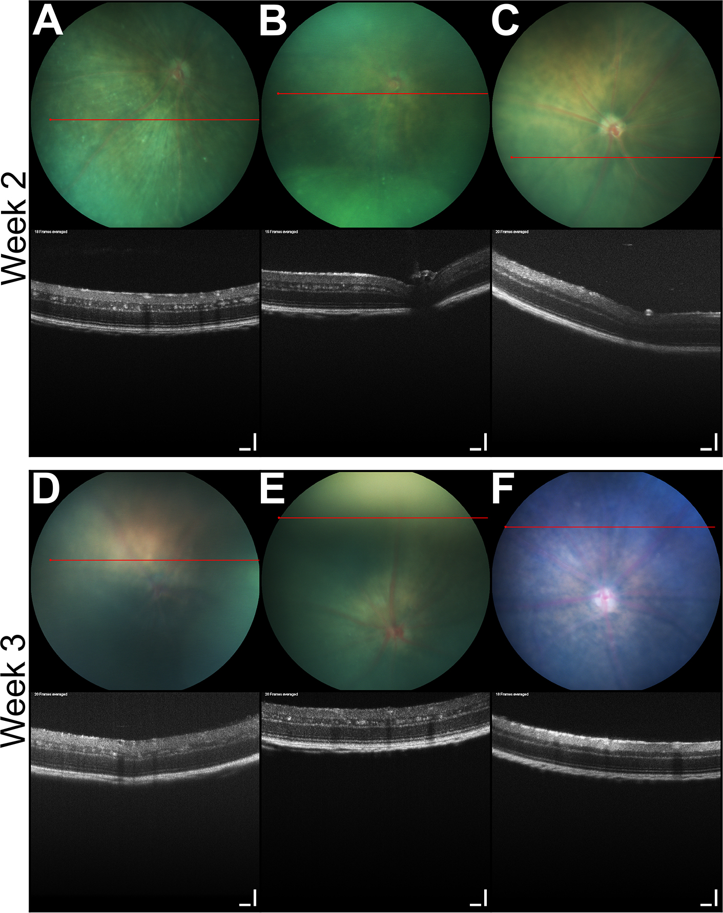
 Figure 1 of
Wenzel, Mol Vis 2015; 21:515-522.
Figure 1 of
Wenzel, Mol Vis 2015; 21:515-522.