Figure 5. Effect of SAHA on keratocyte phenotypes in compressed collagen matrices. Maximum intensity projection images of f-actin organization
in corneal keratocytes following 4 days of culture within compressed collagen matrices. A: Keratocytes in compressed collagen matrices cultured in basal serum-free media (S-) with vehicle developed a dendritic morphology
with membrane-associated f-actin labeling, and this morphology was maintained in the presence of SAHA (B). C: PDGF BB induced keratocyte spreading in compressed collagen matrices without inducing stress fiber formation, and this spreading
response was maintained in the presence of SAHA (D). E: TGFβ1 induced the loss of dendritic processes and the formation of stress fibers, and this transformation was blocked by
SAHA (F).
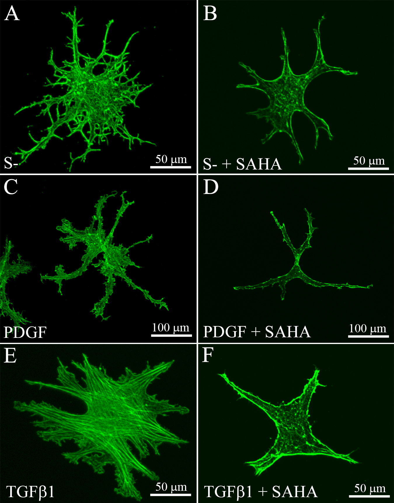
 Figure 5 of
Koppaka, Mol Vis 2015; 21:502-514.
Figure 5 of
Koppaka, Mol Vis 2015; 21:502-514.