Figure 4. Effect of SAHA on cell-induced matrix reorganization. A-F: Maximum intensity projection images of corneal keratocytes following 4 days of culture within 3-D collagen matrices. Green:
f-actin; red: collagen. A: Keratocytes in basal serum-free media (S-) with vehicle have a dendritic morphology and a membrane-associated f-actin distribution,
and they do not induce a significant compaction of collagen ECM. B: Keratocytes in basal media containing SAHA have a similar phenotype. C: PDGF BB induced the formation and elongation of dendritic cell processes without inducing stress fiber formation, and this
spreading response was maintained in the presence of SAHA (D). E: TGFβ1 induces a contractile phenotype, as indicated by a loss of dendritic cell processes, the development of stress fibers,
and local matrix compaction. F: Cells cultured in TGFβ1 plus SAHA are elongated and do not form stress fibers or induce ECM compaction.
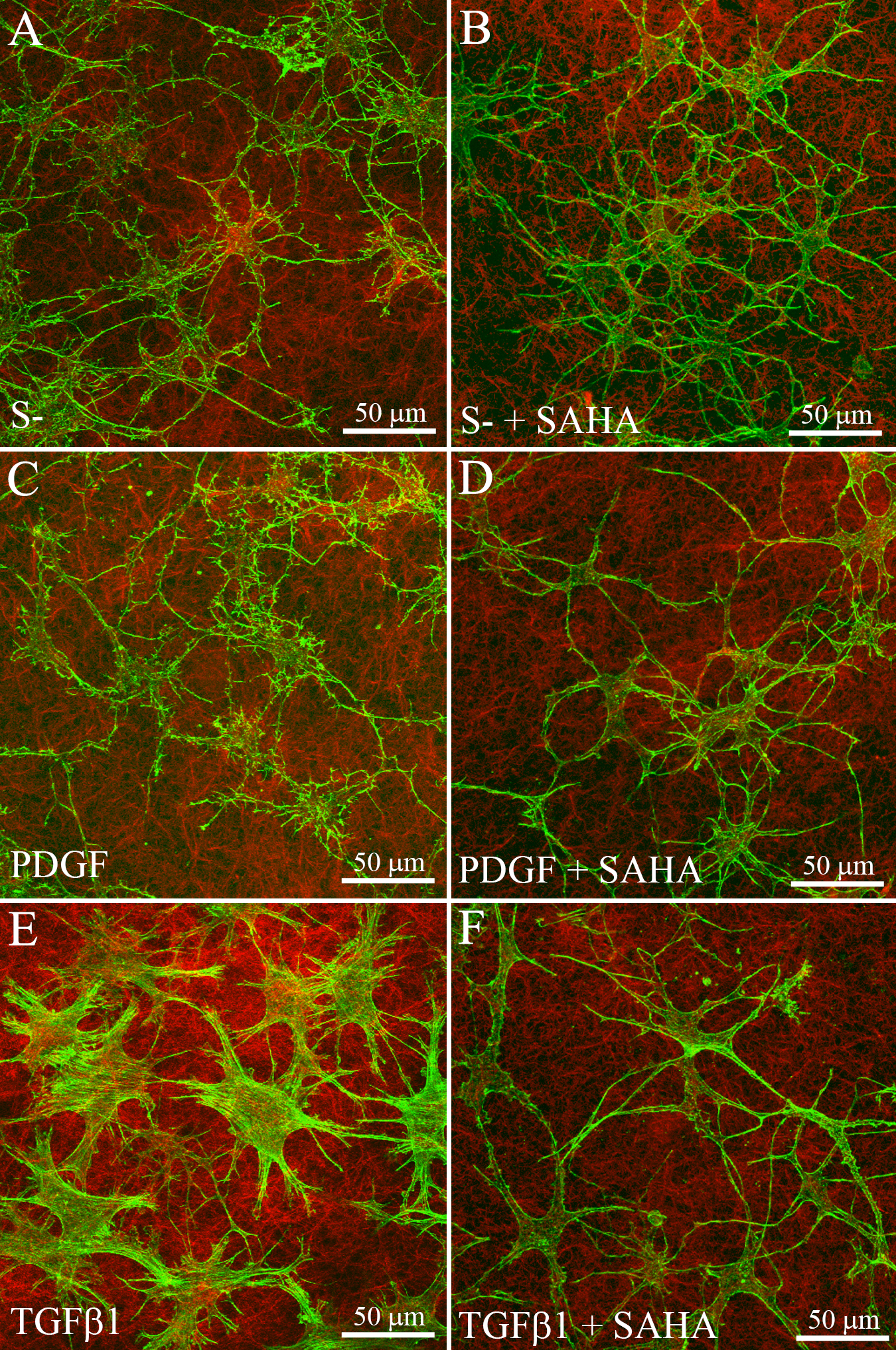
 Figure 4 of
Koppaka, Mol Vis 2015; 21:502-514.
Figure 4 of
Koppaka, Mol Vis 2015; 21:502-514.