Figure 4. SD-OCT in patients with X-linked retinoschisis. A: Patient B,II-1 (18 years) had lamellar schisis. B: Patient C,II-1 (23 years) had photoreceptor and RPE atrophy in the fovea. C: Patient N,II-1 (51 years) had parafoveal schisis and foveal atrophy. D: Patient O,II-1 (30 years) had foveolamellar schisis. E: Patient J,II-2 (5 years) had foveolamellar schisis and lamellar hole. F: Patient K,II-1 (22 years; OS) had foveolamellar schisis and lamellar hole. G: Patient E,II-1 (6 years) had foveal schisis and increased thickness in the fovea. H: Patient M,II-3 (4 years) had foveolamellar schisis and increased thickness in the macula area.
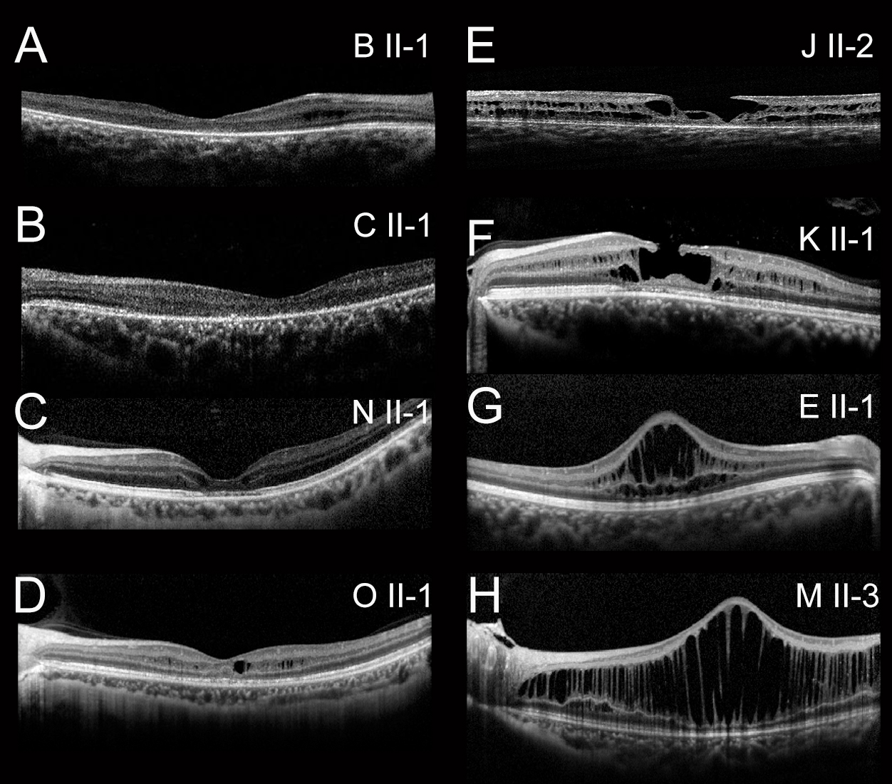
 Figure 4 of
Wang, Mol Vis 2015; 21:487-501.
Figure 4 of
Wang, Mol Vis 2015; 21:487-501.