Figure 3. Fundus autofluorescence (FAF) and non-leakage cystoid macula edema in patients with X-linked retinoschisis. A: FAF for patient A,II-1 (58 years) showed a spoke-wheel pattern of hyper- and hypo-FAF in the macula area. B: Patient C,II-1 (23 years) had irregularly shaped areas of hyper- and hypo-FAF. C: Patient K,II-1 (22 years) had concentric areas of hyper-FAF. D: Patient N,II-1 (51 years) had concentric areas of hyper-FAF (upper), and fluorescence angiography (middle) revealed no leakage
in the central macula, which corresponded to cystoid macula edema on spectral domain optical coherent tomography (bottom).
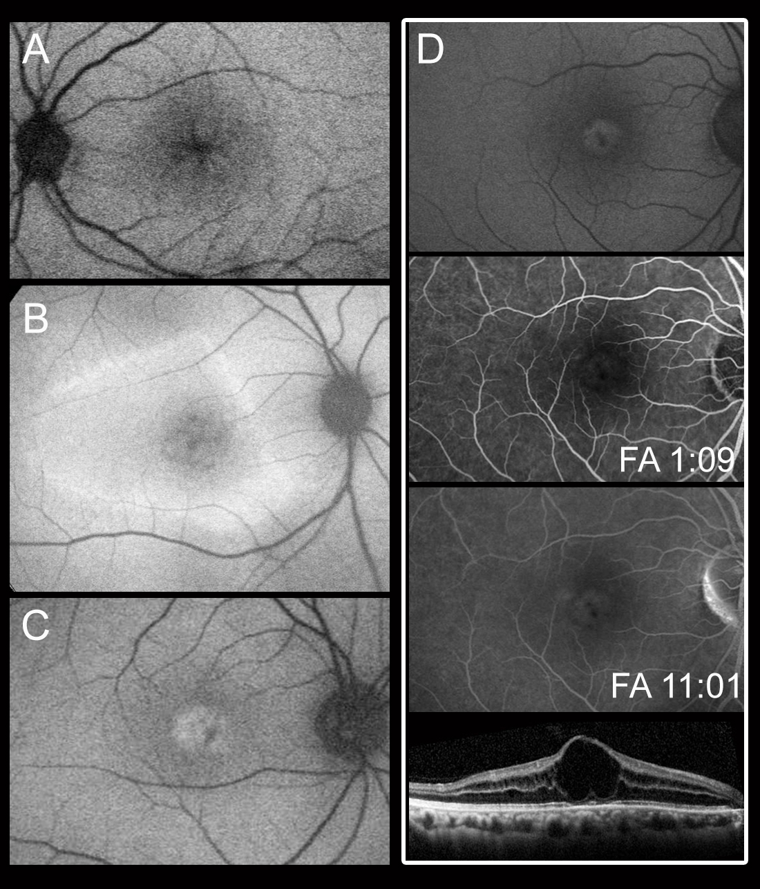
 Figure 3 of
Wang, Mol Vis 2015; 21:487-501.
Figure 3 of
Wang, Mol Vis 2015; 21:487-501.