Figure 6. A section of stained RPE cells. The “before” picture was acquired through the EZ-C1 software accompanying the Nikon microscope
and is displayed without any image processing. The “after” picture was processed using the ProcessImage action script, created for Adobe Photoshop CS3 and included in Appendix 2. The script’s function was to increase the intensity
of the green border staining relative to the background noise found within the cells and balance illumination across the image.
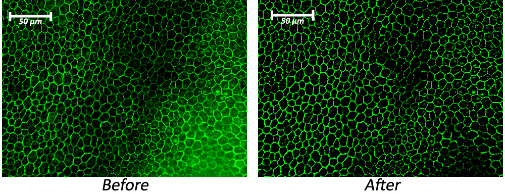
 Figure 6 of
Boatright, Mol Vis 2015; 21:40-60.
Figure 6 of
Boatright, Mol Vis 2015; 21:40-60.